3D Bioprinted Intrahepatic Cholangiocarcinoma Model and 7-Day Perfusion with Gemcitabine and Cisplatin: Modeling the Tumor Microenvironment and Response to Chemotherapy
1Division of Transplant Surgery - Indiana University, Indianapolis, IN, 23D Bioprinting Core and Department of Radiology and Imaging Sciences - Indiana University, Indianapolis, IN, 3Division of Gastroenterology, Richard L. Roudebush VA Medical Center and Indiana University, Indianapolis, IN
Meeting: 2020 American Transplant Congress
Abstract number: D-307
Keywords: Apoptosis, Bile duct, Bioengineering, Liver
Session Information
Session Name: Poster Session D: Cellular Therapies, Tissue Engineering / Regenerative Medicine
Session Type: Poster Session
Date: Saturday, May 30, 2020
Session Time: 3:15pm-4:00pm
 Presentation Time: 3:30pm-4:00pm
Location: Virtual
*Purpose: Over 8,000 patients are newly diagnosed with cholangiocarcinoma (CCA) in the United States each year.Understanding the CCA microenvironment will facilitate the development of novel therapeutic strategies and drug regimens. 3D-bioprinting is useful for studying cell-cell interaction and pathophysiological functions, with advantages over traditional 2D cell culture. We developed and tested a novelscaffold-free3D-bioprinted intrahepatic CCA model, which was continuously perfused with gemcitabine and cisplatin to elucidate tumor-support cell interactions in the setting of chemotherapy.
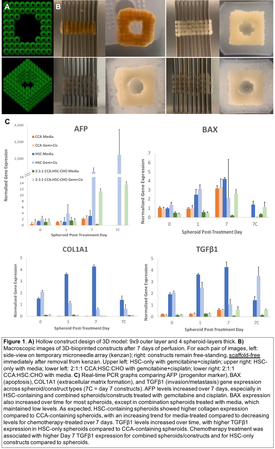
*Methods: All humanprimary intrahepatic CCA (HuCC-T1), hepatic stellate cells (HSC), and cholangiocytes (CHO) were co-cultured to form spheroids. Spheroids were bioprinted using the Regenova 3D-Bioprinter into liver tissue constructs, as HSC-only (control) or CCA:HSC:CHO (2:1:1) combination. Constructs were perfused continuously for 7 days with media-alone or media-supplemented-with-gemcitabine-and-cisplatin (10 μM/L and 20 μM/L, respectively) in the FABRICA bioreactor. Real-time PCR was used to analyze the transcription of AFP, BAX, COL1A1, TGFβ1 for functionality of cancer/progenitor cell markers, apoptosis, extracellular matrix (ECM) deposition, and metastatic potential in spheroids/constructs on days 0, 1, 7.
*Results: The 3D-CCA model was successfully bioprinted and perfused with media-alone or media-gemcitabine/cisplatin for 7 days. Gemcitabine and cisplatin prevented ECM formation and robust tissue fusion in chemotherapy-treated constructs (Figure 1B). Overall, AFP, BAX, COL1A1, TGFβ1 gene expression increased over the treatment period (Figure 1C). Gemcitabine/cisplatin treatment was associated at day 7 with markedly elevated AFP expression, decreasing COL1A1 over time, higher BAX in combined conditions, and higher TGFβ1 expression in HSC-containing constructs compared to spheroids.
*Conclusions: Development of an inexpensive and efficient 3D-CCA model enables us to study its microenvironment for expression of cancer/progenitor markers, apoptosis, collagen formation, and invasion/metastasis under chemotherapy.
To cite this abstract in AMA style:
Chen AM, Zhang W, Sterner J, Kennedy L, Isidan A, Gramelspacher E, Francis HL, Alpini G, Smith LJ, Li P, Ekser B. 3D Bioprinted Intrahepatic Cholangiocarcinoma Model and 7-Day Perfusion with Gemcitabine and Cisplatin: Modeling the Tumor Microenvironment and Response to Chemotherapy [abstract]. Am J Transplant. 2020; 20 (suppl 3). https://atcmeetingabstracts.com/abstract/3d-bioprinted-intrahepatic-cholangiocarcinoma-model-and-7-day-perfusion-with-gemcitabine-and-cisplatin-modeling-the-tumor-microenvironment-and-response-to-chemotherapy/. Accessed February 15, 2026.« Back to 2020 American Transplant Congress

