Understanding the Response of Human Lung Microvascular Endothelial Cells During Lung Transplantation via an In Vitro Model of Ischemia Reperfusion Injury
1Regenerative Medicine Research, Texas Heart Institute, Houston, TX, 2Baylor College of Medicine - Lung Transplant, Houston
Meeting: 2022 American Transplant Congress
Abstract number: 676
Keywords: Apoptosis, Endothelial cells, Ischemia, Lung transplantation
Topic: Basic Science » Basic Science » 14 - Ischemia Reperfusion
Session Information
Session Time: 5:30pm-7:00pm
 Presentation Time: 5:30pm-7:00pm
Location: Hynes Halls C & D
*Purpose: Lung Transplantation (LTx) is a therapeutic option for patients with end-stage lung disease. One of the major complications is primary graft dysfunction largely attributed to ischemia-Reperfusion Injury (IRI) following transplantation. However, our current understanding of IRI mechanisms during lung preservation and transplantation is mainly based on clinical and animal studies. Herein, we aim to develop an in-vitro model to explore the mechanisms of IRI at the endothelial level, which is crucial for maintaining essential lung structure and function.
*Methods: Human Lung Microvascular Endothelial Cells (HLMVCs) line was obtained from Applications, INC. 540-05A. HLMVCs were cultured following provider protocol. IRI model for LTx: cold ischemia was simulated by replacing the cell culture medium with Perfadex® solution and storing the chamber at 4°C for 6h with a gas mixture (4.97 carbon dioxide, 19.6% Oxygen, balanced nitrogen). To simulate reperfusion, the Perfadex® was changed with a warm cell culture medium; cells were then cultured for 24h. The control plates were kept in incubation at 37°C, 5% CO2. Caspase-3/7 was measured by an Apo-One Homogeneous Caspase-3/7 assay, membrane integrity by CellTox, and quantification of lactate dehydrogenase (Promega). CellROX® (Invitrogen) for oxidative stress and DAPI for DNA staining.
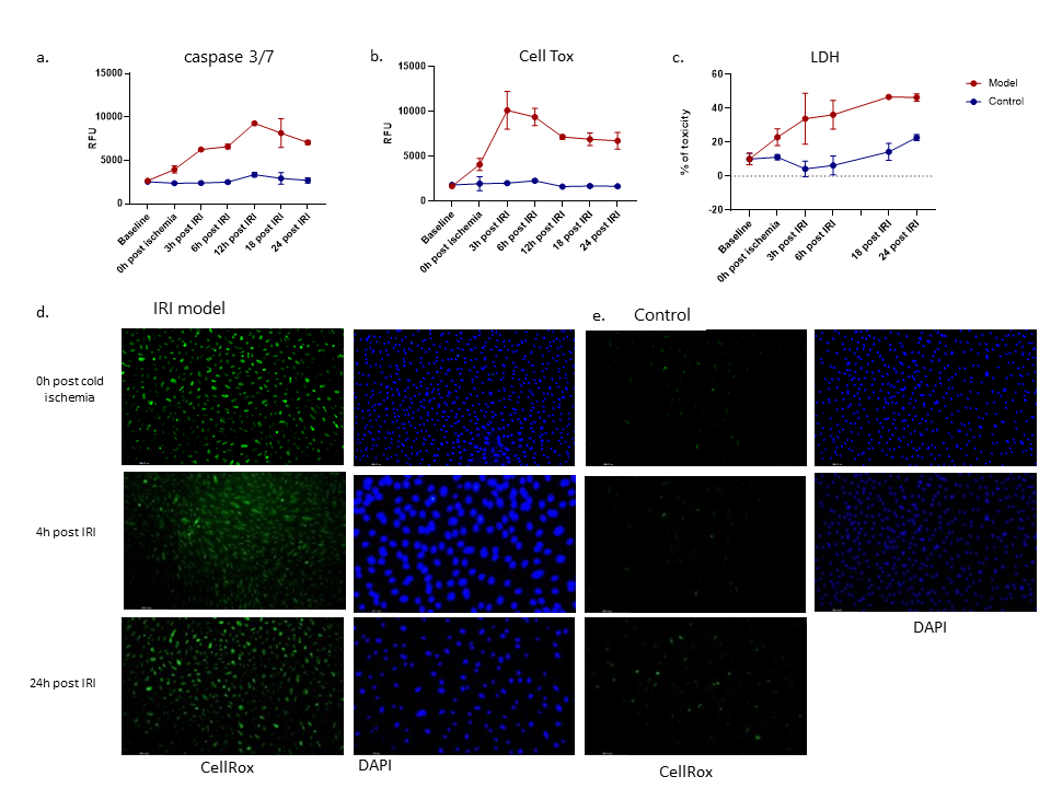
*Results: IRI showed a significant increase of caspase 3/7 (Fig.1a), disruption of membrane integrity(fig.1b) and increased percentage of cell toxicity(LDH) (fig. 1c) after 6h of cold ischemia (0h post-ischemia), with a higher increment during the reperfusion time and it persisted during the next 24h after IRI. (Fig,1d) shows important concentrations of oxidative stress that are present after IRI. These changes were not observed in the control groups (Fig.1e)
*Conclusions: In this study, we showed that HLMVECs suffer significant damage during IRI after LTx. Our findings could contribute to understanding the pathophysiology of PGD and elucidate potential therapeutic targets.
To cite this abstract in AMA style:
Alberty LChacon, King M, Loor G, Curty E, Hochman-Mendez C. Understanding the Response of Human Lung Microvascular Endothelial Cells During Lung Transplantation via an In Vitro Model of Ischemia Reperfusion Injury [abstract]. Am J Transplant. 2022; 22 (suppl 3). https://atcmeetingabstracts.com/abstract/understanding-the-response-of-human-lung-microvascular-endothelial-cells-during-lung-transplantation-via-an-in-vitro-model-of-ischemia-reperfusion-injury/. Accessed February 22, 2026.« Back to 2022 American Transplant Congress

