Inflammasome, Unfolded Protein Response, and T Cell Immunity in DAIH.
1Ped Gastro. and Hep., Yale University, New Haven, CT
2Gastro., Hep., and Nutr., Hospital for Sick Children, Toronto, ON, Canada
3Ped. Gastro., Hep., and Nutr., Columbia University, New York, NY
Meeting: 2017 American Transplant Congress
Abstract number: 565
Keywords: Autoimmunity, Graft survival, Inflammation, Liver transplantation
Session Information
Session Time: 8:00am-10:00am
 Presentation Time: 9:45am-10:00am
Location: Arie Crown Theater
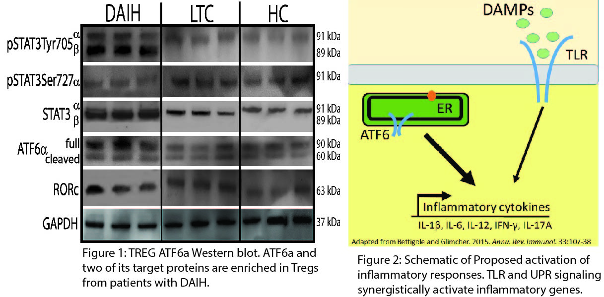
Background: Regulatory T cells (Tregs) of patients with de novo autoimmune hepatitis (DAIH) secrete IL-17A & IFN-g; display TH1 & TH17 pehnotypes & are functionally impaired; monocytes drive differentiation of these Tregs towards TH1-like Tregs in an IL-12 dependent manner. Aim: Identify molecular mechanisms through which Tregs of patients with DAIH differentiate toward pro-inflammatory phenotypes, via both cell-autonomous and non-cell-autonomous pathways. Methods: Blood was obtained from liver transplanted (LT) recipients with DAIH (n=6), LT recipients without DAIH who have normal graft function (LTC) (n=16), & healthy non-transplanted children (HC) (n=13). 1) LPS stimulated CD14+ monocytes were: (i) stained for intracellular IL-1β; (ii) RNA harvested for qRT-PCR for pro-IL-1β; (iii) protein harvested for western blot of inflammasome-associated proteins. 2) DNA harvested from sera for measurement of damage associated molecular patterns (DAMPs) by qRT-PCR. 3) ELISA performed on sera to measure TLR-specific DAMPs. 4) Tregs were FACS isolated and stimulated with PMA/ionomycin for 4-hrs &: (i) total RNA harvested for RNASeq & qPCR for ATF6, RORC, and STAT3; (ii) total protein harvested for western blot of same targets. Comparison of quantitative variables between 2 groups performed using Wilcoxon rank-sum test. P<0.05 statistically significant. Results: In patients with DAIH compared to HC and LTC: CD14+ monocytes display: 1) increased inflammasome activation upon LPS-stimulation (p<0.05), 2) increased pro-IL-1β & IL-1β (p<0.02) (p<0.03) respectively; Increased DAMPs in sera (qPCR p<0.005;ELISA p<0.05); and activation of ATF6 in Tregs as well as two of its targets, RORC & STAT3 (qPCR p<0.01; western blot; cleaved ATF6a p<0.05, RORC p<0.01, and phosphorylated STAT3 p<0.001, Figure 1).Conclusions: Endoplasmic reticulum stress synergizes with the innate immune signaling pathway, TLR, leading to cytokine production (Figure 2). This may explain the pro-inflammatory cytokine secretion observed in Tregs of patients with DAIH. 
CITATION INFORMATION: Arterbery A, Martinez M, Lobritto S, Avitzur Y, Ekong U. Inflammasome, Unfolded Protein Response, and T Cell Immunity in DAIH. Am J Transplant. 2017;17 (suppl 3).
To cite this abstract in AMA style:
Arterbery A, Martinez M, Lobritto S, Avitzur Y, Ekong U. Inflammasome, Unfolded Protein Response, and T Cell Immunity in DAIH. [abstract]. Am J Transplant. 2017; 17 (suppl 3). https://atcmeetingabstracts.com/abstract/inflammasome-unfolded-protein-response-and-t-cell-immunity-in-daih/. Accessed February 11, 2026.« Back to 2017 American Transplant Congress
