Indocyanine Green Fluorescence Imaging during Normothermic Ex Vivo Liver Perfusion for the Assessment of Hepatocyte, Biliary, and Vascular Function
Multi-Organ Transplant Program, Department of Surgery, Toronto General Hospital, Toronto, ON, Canada
Meeting: 2019 American Transplant Congress
Abstract number: A90
Keywords: Image analysis, Liver grafts, Liver metabolism, Liver preservation
Session Information
Session Name: Poster Session A: Ischemia Reperfusion & Organ Rehabilition
Session Type: Poster Session
Date: Saturday, June 1, 2019
Session Time: 5:30pm-7:30pm
 Presentation Time: 5:30pm-7:30pm
Location: Hall C & D
*Purpose: Normothermic Ex Vivo Liver Perfusion (NEVLP) is a novel preservation for the storage and assessment of marginal grafts, such as Donation after Cardio-circulatory Death (DCD). However, accurate parameters reflecting graft injury, graft function, and vascular injury during NEVLP are lacking. Indocyanine Green (ICG) is a substance that is exclusively cleared by hepatocytes and excreted into the bile without biotransformation. Furthermore, ICG emits fluorescence when excited by near-infrared light and various studies have validated the use of ICG imaging in operation. We evaluated arterial perfusion of DCD grafts during NEVLP and determined uptake and excretion of ICG by fluorescence imaging.
*Methods: Thirty to 35 kg male Yorkshire pigs were allocated to 3 groups; Heart beating donor (HBD), DCD 60minutes (moderate damage) and DCD 120 minutes (severe damage), each n = 1. Following induction of warm ischemia with heparinization, procurement and 2-hour cold storage, the livers were connected to the NEVLP circuit. We measured liver fluorescence for 4 hours after ICG injection via artery with clamping portal vein for 1 minute. Regional fluorescence data was drawn from SPY Elite® System (Stryker) and analyzed by Image J (National Institutes of Health). To quantify the arterial ischemic area, the rates of low intensity areas within regions were calculated. The threshold of intensity was 100; that is approximately 40% of Maximum intensity.
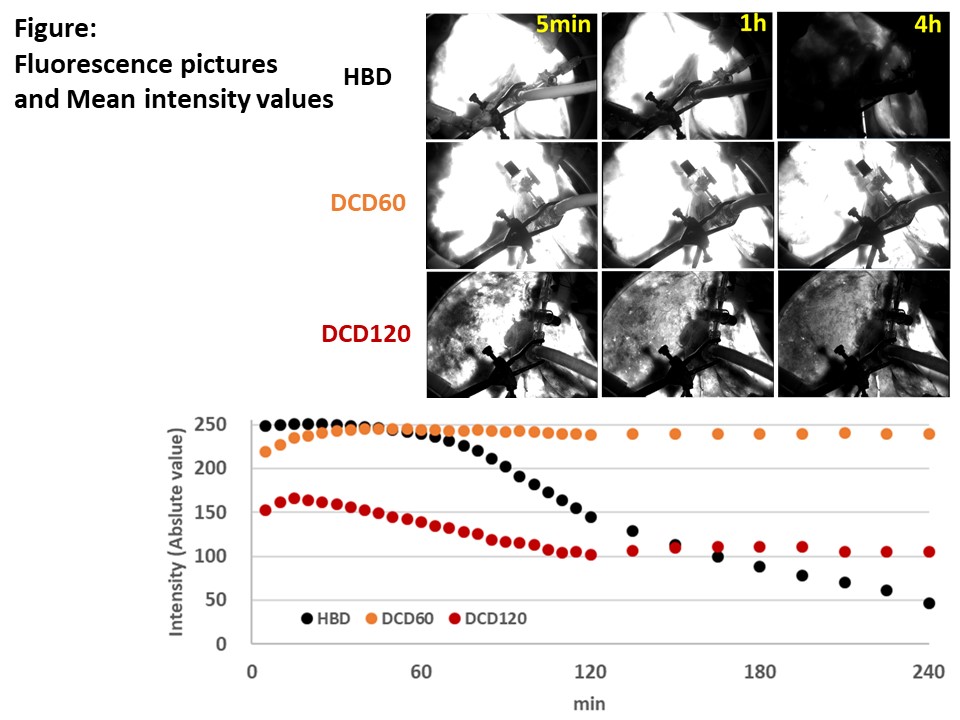
*Results: In the early phase after arterial ICG injection during the clamping of the portal vein, fluorescence imaging demonstrated low parenchyma perfusion in DCD livers with prolonged ischemia. The low intensity rate, as a marker of poor perfusion, was higher with longer warm ischemia times; HBD 1.1%, DCD 60minutes 18.9%, and DCD 120minutes 37.5%. HBD liver showed homogeneous fluorescence and ICG was excreted quickly into the bile. In the DCD 60minutes group the liver demonstrated low perfusion areas in peripheral liver parenchyma and the high intensity had remained for 4 hours. The DCD 120minutes liver had worse perfusion without central area perfusion and the intensity remained high without clearance after NEVLP. (Figure)
*Conclusions: Our preliminary results indicate that ICG imaging during NEVLP reflects the severity of ischemic injury and might provide useful information for graft hepatocyte, biliary and vascular function.
To cite this abstract in AMA style:
Goto T, Linares I, Ganesh S, Mazilescu L, Urbanellis P, Selzner N, Selzner M. Indocyanine Green Fluorescence Imaging during Normothermic Ex Vivo Liver Perfusion for the Assessment of Hepatocyte, Biliary, and Vascular Function [abstract]. Am J Transplant. 2019; 19 (suppl 3). https://atcmeetingabstracts.com/abstract/indocyanine-green-fluorescence-imaging-during-normothermic-ex-vivo-liver-perfusion-for-the-assessment-of-hepatocyte-biliary-and-vascular-function/. Accessed May 21, 2026.« Back to 2019 American Transplant Congress