PPAR-γ Agonist Attenuates Hepatic Ischemia-Reperfusion Injury in a Mouse Model by Regulating Kupffer Cell Polarization.
Multi-Organ Transplant Program, Toronto General Hospital, University Health Network, Toronto, ON, Canada
Meeting: 2017 American Transplant Congress
Abstract number: D50
Session Information
Session Name: Poster Session D: Ischemic Injury and Organ Preservation Session III
Session Type: Poster Session
Date: Tuesday, May 2, 2017
Session Time: 6:00pm-7:00pm
 Presentation Time: 6:00pm-7:00pm
Location: Hall D1
PPAR-γ agonists have shown protective effect on ischemia-reperfusion injury (IRI) in cerebral and renal tissue. The effects of PPAR-γ on hepatic IRI is unclear. We investigated the effects and mechanisms of protection of PPAR-γ agonists on hepatic IRI.
Methods:
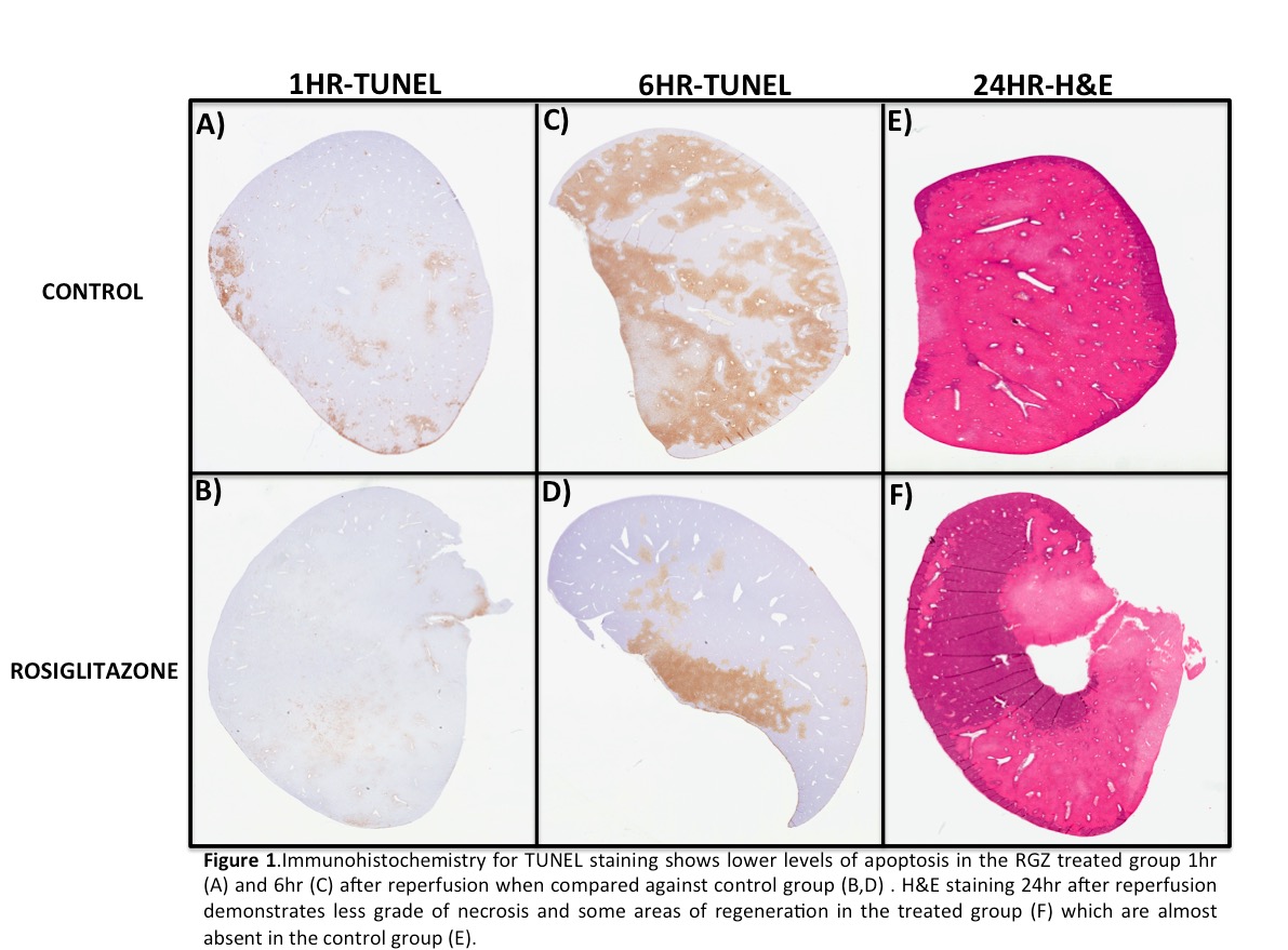
Ischemia of 70% of the liver was induced for 60min. PPAR-γ agonist Rosiglitazone (RGZ-3mg/kg) or vehicle (control) were administrated 24 and 1hr before reperfusion. Aspartate aminotransferase (AST) was measured 1, 6, 12 and 24hrs after reperfusion as a marker of hepatocyte injury. H&E and TUNEL staining was performed to evaluate hepatic necrosis and apoptosis, respectively. Flow-cytometry was used to assess Kupffer cells polarization (M2-CD206+, M1-Nitric Oxide +).
Results:
AST in the RGZ group presented significant reduction after 1hr (RGZ: 3092±279 vs Control: 4469±1559, p=0.042), 6hr (RGZ: 7041±3281 vs Control: 12193±4329, p=0.015), and 12hr (RGZ: 5746±868 vs Control: 8608±3560, p=0.049) of reperfusion. TUNEL staining was significantly reduced in the RGZ vs control group at 1hr (RGZ: 2.46±1.47 vs Control: 6.90±2.42, p=0.001) and 6hrs (RGZ: 28±22 vs Control: 54±20, p=0.027) after reperfusion. H&E staining demonstrated a decreased percentage of necrotic tissue in the treated group 24hr (RGZ: 59±15 vs Control 76±12) after reperfusion. Flow-cytometry revealed that previous to the ischemic insult the percentage of M2 Kupffer cells was increased in the RGZ vs control group (RGZ: 9.6±0.7 vs Control: 4.8% ±0.8, p=0.04). Percentage of M1-Kupffer cells polarization was diminished in the treated group when compared with the control group since 1hr after reperfusion (RGZ: 23±10 vs Control: 45±22, p=0.083), this difference became significant 6hs (RGZ: 3.93±3 vs Control: 31±21, p=0.025) and 24hs (RGZ: 4.15±3 vs Control: 17.10±9, p=0.043) following reperfusion. Conclusion:
PPAR-γ reduces hepatic IRI injury and decreases M1 polarization of Kupffer cells. Kupffer cell polarization is a novel target to reduce hepatic reperfusion injury.
CITATION INFORMATION: Linares I, Farrokhi K, Echeverri J, Kaths M, Kollman D, Hamar M, Urbanellis P, Ganesh S, Adeyi O, Yip P, Selzner N, Selzner M. PPAR-γ Agonist Attenuates Hepatic Ischemia-Reperfusion Injury in a Mouse Model by Regulating Kupffer Cell Polarization. Am J Transplant. 2017;17 (suppl 3).
To cite this abstract in AMA style:
Linares I, Farrokhi K, Echeverri J, Kaths M, Kollman D, Hamar M, Urbanellis P, Ganesh S, Adeyi O, Yip P, Selzner N, Selzner M. PPAR-γ Agonist Attenuates Hepatic Ischemia-Reperfusion Injury in a Mouse Model by Regulating Kupffer Cell Polarization. [abstract]. Am J Transplant. 2017; 17 (suppl 3). https://atcmeetingabstracts.com/abstract/ppar-agonist-attenuates-hepatic-ischemia-reperfusion-injury-in-a-mouse-model-by-regulating-kupffer-cell-polarization/. Accessed February 10, 2026.« Back to 2017 American Transplant Congress
