Pathological Characteristics of BK Polyomavirus Nephropathy with Glomerular Involvement
1Department of Organ Transplantation, 1st Affiliated Hospital, Zhongshan University, Guangzhou, China, 2Department of Pathology, 1st Affiliated Hospital, Zhongshan University, Guangzhou, China
Meeting: 2020 American Transplant Congress
Abstract number: A-214
Keywords: Biopsy, Glomerulonephritis, Kidney/liver transplantation, Polyma virus
Session Information
Session Name: Poster Session A: Kidney: Polyoma
Session Type: Poster Session
Date: Saturday, May 30, 2020
Session Time: 3:15pm-4:00pm
 Presentation Time: 3:30pm-4:00pm
Location: Virtual
*Purpose: This study aimed to investigate the pathological characteristics of BK polyomavirus-associated nephropathy with glomerular involvement in kidney transplant recipients.
*Methods: Forty-four patients with glomerular BK polyomavirus (BKPyV) infection were included for analysis. Immunohistochemical (IHC) staining was performed on paraffin sections using monoclonal mouse anti-SV40 large T antigen antibody.
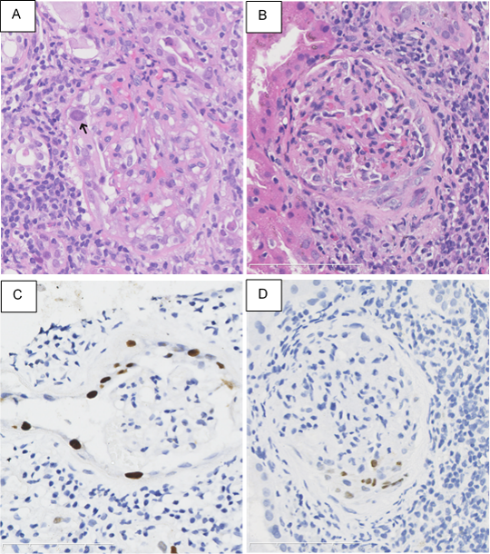
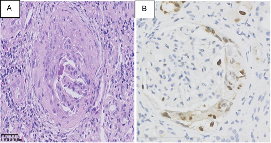
*Results: In BKPyV-infected glomeruli, the glomerular parietal epithelial cells (GPECs) were swollen, hyperchromatic, and enlarged, with an increased nuclear to cytoplasm (N/C) ratio and smudgy basophilic intra-nuclear viral inclusions. IHC staining revealed the distribution of BKPyV involvement in GPECs, podocytes, and shedding cells within Bowman’s space (Figure 1). Banff g1 glomerulitis was found in 43.2% of the biopsies. Mononuclear inflammatory cells were found mainly in the glomerular capillary wall, and occasionally among the proliferating GEPCs. Peri-glomerular fibrosis was the most common chronic change (65.9%). Notably, BKPyV affected GEPC proliferation and caused crescent formation (7 biopsies, 15.9%). Three biopsies exhibited fibrous crescents and the absence of viral inclusions. The other 4 biopsies exhibited cellular and fibro-cellular crescents, with viral cytopathic changes and positive IHC staining in the proliferative GPECs (Figure 2). Electron microscopy showed viral particles in both GPECs and podocytes. Most BKPyV-infected GPECs were degenerative, with mitochondrial swelling, endoplasmic reticulum expansion, and multi-layered membranous structure formation. Twelve (27.3%) patients received repeat biopsies within 1.6 to 39.5 months (median: 13.5 months), but none revealed persistent glomerular BKPyV infection.
*Conclusions: BKPyVAN with glomerular involvement presents distinct glomerular changes.
To cite this abstract in AMA style:
Huang G, Chen X, Chen W, Yang S, Qiu J. Pathological Characteristics of BK Polyomavirus Nephropathy with Glomerular Involvement [abstract]. Am J Transplant. 2020; 20 (suppl 3). https://atcmeetingabstracts.com/abstract/pathological-characteristics-of-bk-polyomavirus-nephropathy-with-glomerular-involvement/. Accessed February 8, 2026.« Back to 2020 American Transplant Congress


