Mononuclear Cell Infiltrates Characterization in Scar Tissue from a Bilateral Forearm Transplant Patient: Findings to Think
1Immunology, INCMNSZ, Mexico City, Mexico
2Plastic Surgery, INCMNSZ, Mexico, Mexico
3Transplants, INCMNSZ, Mexico, Mexico
4Pathology, INCMNSZ, Mexico City, Mexico.
Meeting: 2015 American Transplant Congress
Abstract number: B288
Keywords: CD20, CD4, Mononuclear leukocytes, T helper cells
Session Information
Session Name: Poster Session B: Vascularized Composite Tissue Allografts and Xenotransplantation
Session Type: Poster Session
Date: Sunday, May 3, 2015
Session Time: 5:30pm-6:30pm
 Presentation Time: 5:30pm-6:30pm
Location: Exhibit Hall E
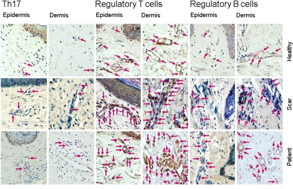
Herein we report the cell infiltrating pattern in scar tissue resected at the junction of the VCA and the tissue corresponding to the recipient arm, at 766 days post-transplant and in the absence of any clinical skin changes suggesting rejection. Immunosuppressive therapy included thymoglobulin induction and initial and maintenance triple therapy based on tacrolimus, MMF and prednisone. The patient previously had 3 acute cellular rejection events: day 60 (grade I); day 385 (grade II); and day 522 posttransplant (grade II) which resolved with treatment. For cell infiltrating pattern comparison, normal skin biopsies (NS, n=5) and hypertrophic scars (HSc, n=5) proceeding from non-VCA patients were analyzed. Methods. Immunohistochemical staining was done in order to determine CD3+, CD4+, CD8+, FOXP3+, CD20+ and CD68+ cells. To determine the subpopulation of CD4+/IL-17A+-expressing T cells (Th17), CD25+/Foxp3+ regulatory T cells (Tregs), and CD20+/IL-10+-producing B cell (Bregs), a simultaneous immunohistochemistry detection was performed. Results. VCA patient scar had dermal infiltrate composed predominantly of T cells (CD3+); both helper (CD4+) and cytotoxic (CD8+), admixed with B cells (CD20+) mainly observed in deep location. Superficial T cells close to basal membrane were observed. Immunohistological analysis showed that VCA patient scar and HSc were similar but different from NS. Surprisingly, no differences in Th17, Tregs, and Bregs percentage were detected between VCA patient scar and HSc. Nonetheless, Tregs and Bregs levels were conspicuously higher in dermis and epidermis (20-30%) vs. NS tissue (5-10%). Th17 cells showed a similar percentage in NS, VCA patient scar and HSc (5%). Conclusions. Characterization of mononuclear cell infiltration in our transplanted patient suggested a preponderance of regulatory cells vs. inducing cells of vasculopathy and rejection (Th17), similar to biopsies of HSc from non-VCA patients. Arrows depict immunoreactive cells.
To cite this abstract in AMA style:
Furuzawa-Carballeda J, Iglesias M, Alberu J, Gamboa A, Butrón P, Morán-Romero M, Esquivel Y. Mononuclear Cell Infiltrates Characterization in Scar Tissue from a Bilateral Forearm Transplant Patient: Findings to Think [abstract]. Am J Transplant. 2015; 15 (suppl 3). https://atcmeetingabstracts.com/abstract/mononuclear-cell-infiltrates-characterization-in-scar-tissue-from-a-bilateral-forearm-transplant-patient-findings-to-think/. Accessed July 12, 2025.« Back to 2015 American Transplant Congress
