Modification of DC Phenotype and Function via Targeted Porous Silicon Nanoparticles.
1School of Medicine, University of Adelaide, Adelaide, Australia
2Future Industries Institute, University of South Australia, Adelaide, Australia
3CNARTS, Royal Adelaide Hospital, Adealide, Australia
4Transplantation Immunology, Garvan Institute of Medical Research, Sydney, Australia.
Meeting: 2016 American Transplant Congress
Abstract number: 204
Keywords: Immunosuppression, Sirolimus (SLR), T cells, Tolerance
Session Information
Session Name: Concurrent Session: Innate Immunity in Rejection and Tolerance: Animal Models
Session Type: Concurrent Session
Date: Monday, June 13, 2016
Session Time: 2:30pm-4:00pm
 Presentation Time: 3:30pm-3:42pm
Location: Room 313
Aims: Dendritic cells (DC) are the most potent antigen-presenting cell and are fundamental in the establishment of transplant tolerance. Targeting DC via the DC-SIGN receptor is a potential target for cell specific therapy. Porous silicon nanoparticles (pSiNP) loaded with immunosuppressant rapamycin (RAPA-pSiNP) provide a unique platform to target and modify DC in vivo. The aim of this study was to conjugate monoclonal antibody anti-DC-SIGN on to the surface of RAPA-pSiNP and determine the effects on targeted DC phenotype and stimulatory capacity in vitro. Methods: Fluorescein isothiocyanate (FITC)-labelled pSiNP conjugated to either anti-DC-SIGN or isotype control were cultured with whole blood samples in vitro to assess specific targeting of DC. Uptake was determined via flow cytometry and transmission electron microscopy.  Rapamycin loading of pSiNP was confirmed with ultraviolet visualisation and inferred spectrometry. DC were co-cultured with rapamycin loaded pSiNP for 2 days (± LPS), irradiated and co-cultured with CFSE stained allogeneic T-cells. Results: Anti-DC-SIGN pSiNP favourably targeted and were phagocytised by myeloid DC in whole blood samples in a time and dose dependent manner. Myeloid DC were 42% positive for Anti-DC-SIGN functionalised NP compared to only 10% for Isotype control and 5% for unfunctionalised NP. DC preconditioning with RAPA-pSiNP results in a maturation resistant phenotype and significantly suppresses allogeneic T-cell proliferation by 28.6 ± 1.9% (p<0.0001). Conclusions: RAPA-pSiNP conjugated to anti-DC-SIGN actively targets and modifies DC function and may serve as a novel therapy to target DC in vivo.
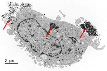
Figure 1: Transmission electron micrograph of human monocyte-derived DC treated with anti-DC-SIGN pSiNP for 2 hours. Arrows show pSiNP within lysosome and bound to surface of DC.
CITATION INFORMATION: Stead S, McInnes S, Kireta S, Rose P, Rojas-Canales D, Jesudason S, Grey S, Carroll R, Voelcker N, Coates P. Modification of DC Phenotype and Function via Targeted Porous Silicon Nanoparticles. Am J Transplant. 2016;16 (suppl 3).
To cite this abstract in AMA style:
Stead S, McInnes S, Kireta S, Rose P, Rojas-Canales D, Jesudason S, Grey S, Carroll R, Voelcker N, Coates P. Modification of DC Phenotype and Function via Targeted Porous Silicon Nanoparticles. [abstract]. Am J Transplant. 2016; 16 (suppl 3). https://atcmeetingabstracts.com/abstract/modification-of-dc-phenotype-and-function-via-targeted-porous-silicon-nanoparticles/. Accessed February 9, 2026.« Back to 2016 American Transplant Congress
