A Novel Microfluidic Device for Purification of Pancreatic Islet Preparations.
1Electrical and Computer Engineering, University of Virginia, Charlottesville, VA
2Surgery, University of Virginia, Charlottesville, VA
3Biomedical Engineering, University of Virginia, Charlottesville, VA.
Meeting: 2016 American Transplant Congress
Abstract number: D52
Keywords: Graft function, Islets, Pancreas, Resource utilization
Session Information
Session Name: Poster Session D: Chimerism/Stem Cells, Cellular/Islet Transplantation, Innate Immunity, Chronic Rejection
Session Type: Poster Session
Date: Tuesday, June 14, 2016
Session Time: 6:00pm-7:00pm
 Presentation Time: 6:00pm-7:00pm
Location: Halls C&D
Purpose: Demonstrate that novel continuous flow microfluidic designs can be utilized to remove contaminating acinar tissue and sort the purified islets, based on size and deformability of the pancreatic tissue spheroids.
Methods: The microfluidic device is constructed out of PDMS using lithographic techniques. Beads of known size and suspended pancreas tissue following modified Ricordi chamber islet isolation and purification are used to validate the function of the device design. Both cross-flow and hydrodynamic filtration methods enable continuous separation of pancreatic islet tissue from acinar tissue.
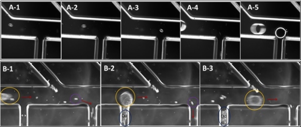
Results:The small size and high deformability acinar tissue pass down the branching channels as the larger and less deformable pancreatic islets are carried out the main outlet by crossflow. High frame rate time lapse photography shows that small particles (35 micron polystyrene bead) exit the main flow (horizontal in Fig A-1 to 3) into one of the separation channels (oriented vertically in the images), while 150 micron beads remain in the main channel (Fig A-4,5). When a pancreatic digest is introduced into the device islets (yellow indicator) are not able to enter the separation channels, while acinar tissue and contaminating particles (blue and violet, respectively) can leave the main flow (Fig B-1 to 3). Variation in applied flow velocity or hydrodynamic resistance of the main channel is used to ensure that large particles do not become lodged at the entrance to separation channels.
 Figure: Time lapse images showing separation of model polystyrene beads (A) and pancreatic tissue (B).
Conclusion: High fidelity separation is achieved by flowing the cell suspension along a series of branching channels. Small particles, such as individual cells, and deformable contaminating acinar tissue are thus separated from the purified islet preparation. Current throughput on this device indicates that it may be utilized for automated quantification of islet preparation purity.
CITATION INFORMATION: Varhue W, Langman L, Bowers D, Peirce-Cottler S, Swami N, Brayman K. A Novel Microfluidic Device for Purification of Pancreatic Islet Preparations. Am J Transplant. 2016;16 (suppl 3).
To cite this abstract in AMA style:
Varhue W, Langman L, Bowers D, Peirce-Cottler S, Swami N, Brayman K. A Novel Microfluidic Device for Purification of Pancreatic Islet Preparations. [abstract]. Am J Transplant. 2016; 16 (suppl 3). https://atcmeetingabstracts.com/abstract/a-novel-microfluidic-device-for-purification-of-pancreatic-islet-preparations/. Accessed February 21, 2026.« Back to 2016 American Transplant Congress
