Microvascular Inflammation in Kidney Transplant Biopsies in the Absence of HLA-DSA Displays Intense Cytotoxic T-cell and NK Cell but Not Plasma Cell Infiltration
1Nephrology Department, Hospital del Mar, Institute Mar for Medical Research (IMIM), Barcelona, Spain, 2Pathology Department, Hospital del Mar, Institute Mar for Medical Research (IMIM), Barcelona, Spain, 3Nephrology Department, Hospital Universitario 12 de Octubre. Institute Mar for Medical Research (IMIM), Madrid, Spain
Meeting: 2022 American Transplant Congress
Abstract number: 573
Keywords: Biopsy, Natural killer cells, Rejection, T cell graft infiltration
Topic: Basic Science » Basic Clinical Science » 19 - Chronic Organ Rejection
Session Information
Session Name: Chronic Organ Rejection
Session Type: Rapid Fire Oral Abstract
Date: Tuesday, June 7, 2022
Session Time: 5:30pm-7:00pm
 Presentation Time: 5:50pm-6:00pm
Location: Hynes Room 309
*Purpose: Microvascular inflammation (MVI) without evidence of HLA-donor-specific antibodies (HLA-DSA) or C4d+ deposition remains an enigmatic phenotype, which cannot be classified as antibody-mediated rejection (ABMR) according to recent Banff classifications.
*Methods: We aimed to compare peripheral blood lymphocyte (PBL) distribution and infiltrating immune cells in kidney transplant (KT) biopsies presenting MVI (g+ptc≥2) without C4d+ and HLA-DSA, ABMR, and normal histology to explore the role of immune cells in these entities.
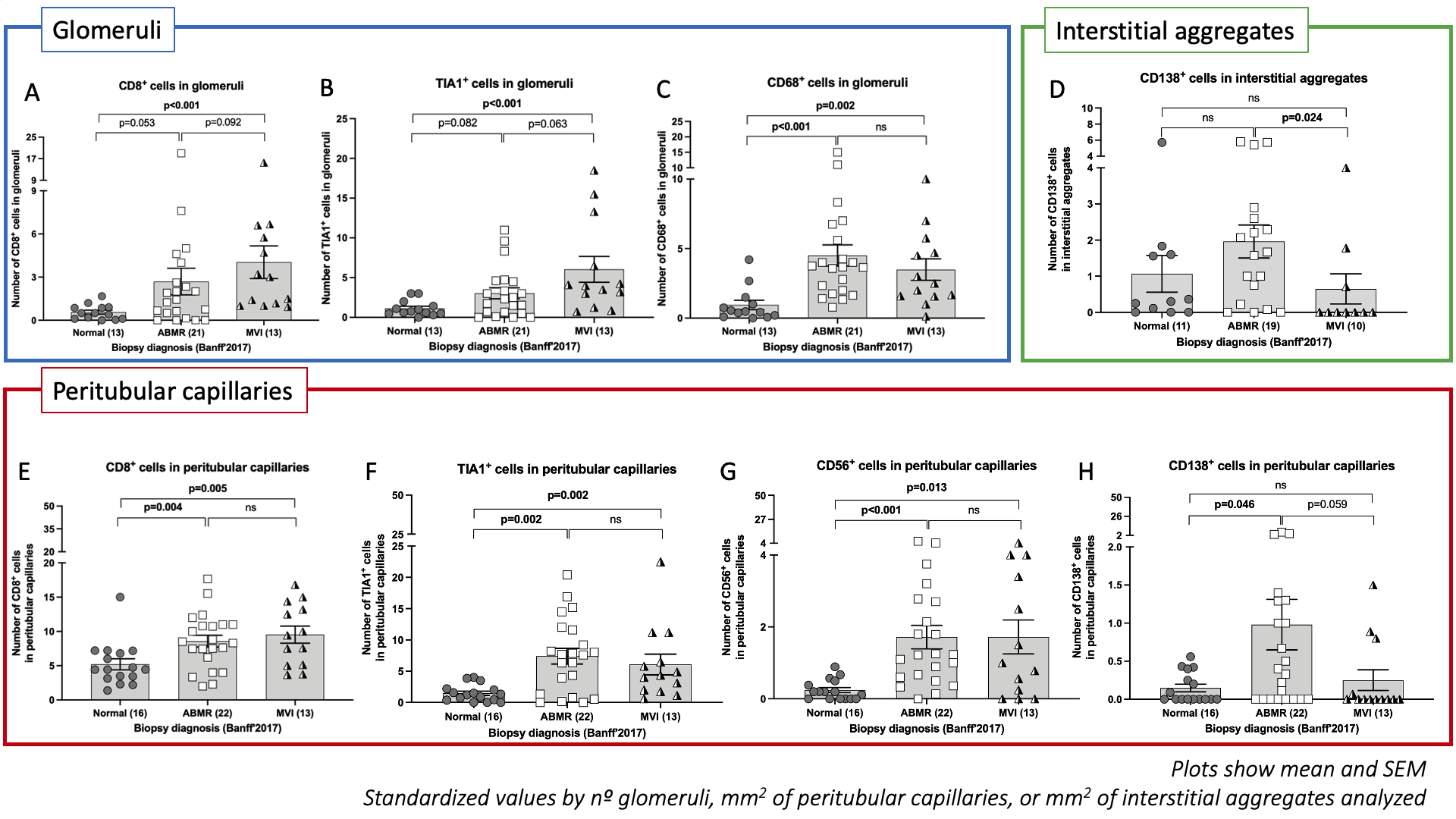
*Results: From 221 allograft biopsies with ABMR (n=73), MVI (n=32), and normal (n=116) diagnosis. MVI patients showed a decrease in the absolute number of T-cells compared with ABMR and normal cases (p=0.020 and p=0.006) due to a significant decrease of CD4+ T-cells compared to normal cases (p=0.013) and a reduction of CD8+ T-cells compared to ABMR (p=0.029). ABMR and MVI presented a lower absolute number of circulating Natural Killer (NK) cells than normal cases. Immunohistochemistry assessment was performed in 22 ABMR, 13 MVI, and 16 normal cases. Glomeruli in ABMR and MVI had more T-cells and CD68+ infiltration than normal biopsies, although TIA1+ was only increased in MVI (p<0.001), suggesting heightened T-cell cytotoxic capacity. Peritubular capillaries displayed more circulating T-cells, CD56+, TIA1+, and CD68+ in ABMR and MVI groups. Contrarily, MVI cases showed mild circulating plasma cell infiltration (CD138+) in peritubular capillaries (p=0.059) and interstitial aggregates (p=0.024) compared to ABMR (Figure).
*Conclusions: In conclusion, MVI without HLA-DSA and C4d+ displays decreased circulating T-cell and NK cells, and intense T-cell and NK cell cytotoxic infiltration in the allograft, similar to ABMR. However, the deficiency of plasma cell infiltration in MVI suggests a different underlying stimulus than ABMR.
Figure. Immunohistochemical assessment in KT biopsies.
To cite this abstract in AMA style:
Buxeda A, Llinàs-Mallol L, Gimeno J, Redondo-Pachón D, Arias-Cabrales C, Burballa C, Puche A, Pérez-Sáez M, Pascual J, Crespo M. Microvascular Inflammation in Kidney Transplant Biopsies in the Absence of HLA-DSA Displays Intense Cytotoxic T-cell and NK Cell but Not Plasma Cell Infiltration [abstract]. Am J Transplant. 2022; 22 (suppl 3). https://atcmeetingabstracts.com/abstract/microvascular-inflammation-in-kidney-transplant-biopsies-in-the-absence-of-hla-dsa-displays-intense-cytotoxic-t-cell-and-nk-cell-but-not-plasma-cell-infiltration/. Accessed February 16, 2026.« Back to 2022 American Transplant Congress

