Non-Invasive Detection of Rejection with ImmunoPET Imaging.
1Department of Surgery, Emory University, Atlanta, GA
2Department of Biomedical Engineering, Georgia Tech, Atlanta, GA
3JN Biosciences LLC, Mountain View, CA
Meeting: 2017 American Transplant Congress
Abstract number: 189
Keywords: Co-stimulation, Non-invasive diagnosis, Primates, T cell graft infiltration
Session Information
Session Name: Concurrent Session: T Cell Biology and Alloimmunity
Session Type: Concurrent Session
Date: Sunday, April 30, 2017
Session Time: 4:30pm-6:00pm
 Presentation Time: 4:54pm-5:06pm
Location: E352
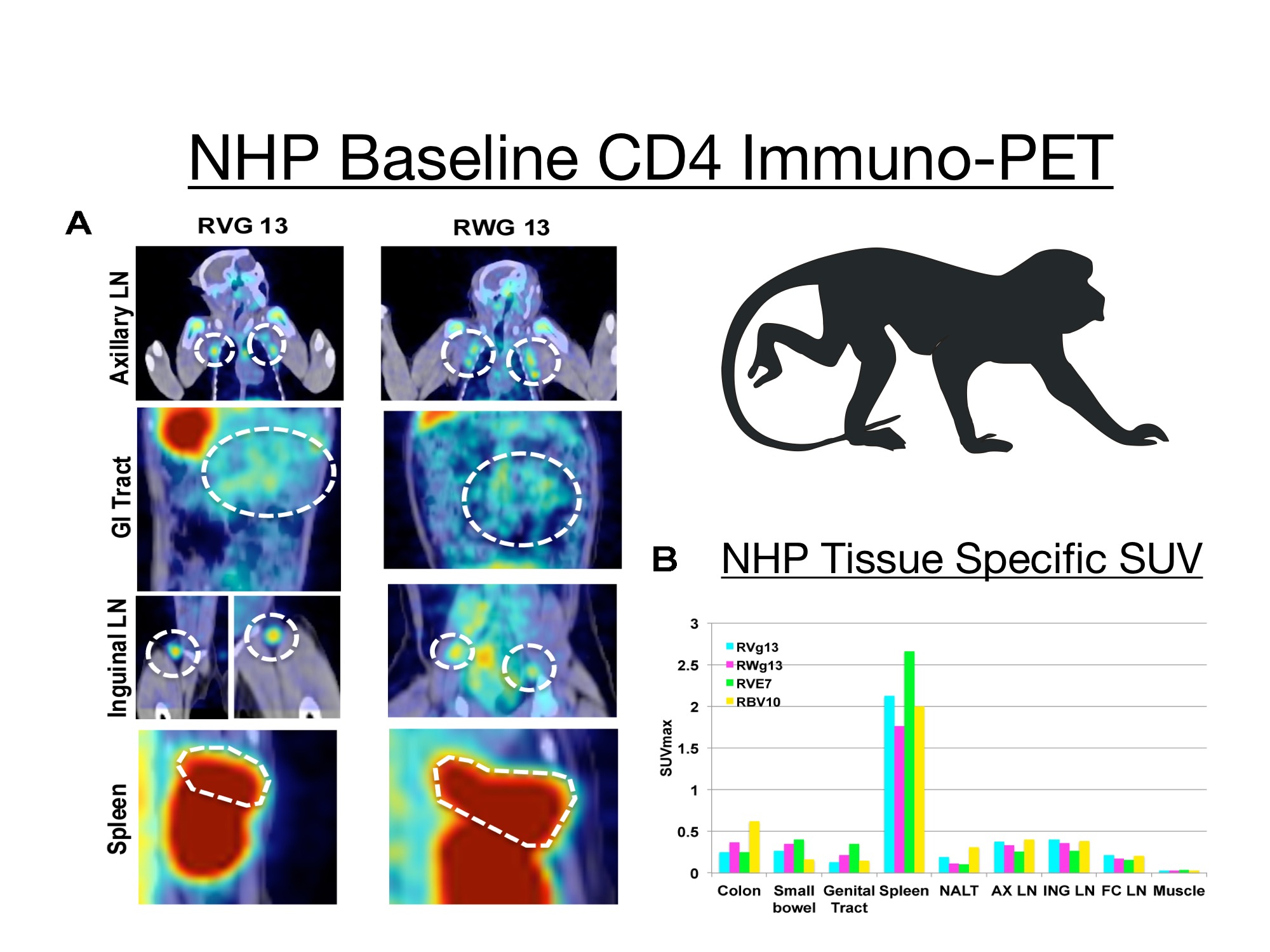
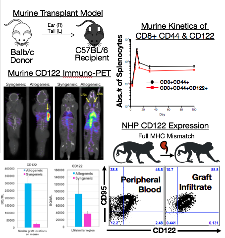
Current approaches to detect and diagnose rejection are retrospective and/or invasive. These methods assess the extent of end organ damage by insensitive and non-specific measures (e.g. Creatinine), or assess graft immune cell infiltrate and organ damage by costly and risky invasive biopsy. The need for early non-invasive detection of alloreactivity prior to organ damage has never been greater. Recently we reported development of a technique to track immune cell subsets during viral infection and T cell depletion in rhesus macaques (Science 2016, Nature Methods 2015). This technique, Immuno-PET (Positron Emission Tomography), allows for whole-body visualization of immune cell subsets with radiolabeled monoclonal antibody or fragments (mAbs) specific for cell surface markers such as CD4. We adapted ImmunoPET for detection of alloreactive T cells. PET is a clinical mainstay for the detection of cancer, using radiolabeled probes to monitor target tissues. We recently described a critical role for CD122 the shared IL2/IL15Rβ, in alloreactivity. CD122 is highly expressed on alloreactive T cells during rejection. T cell infiltrate within the allograft expresses high levels of CD122. We demonstrate the utility of ImmunoPET to sensitively and specifically monitor whole T cell distribution in both mice and non-human primates, and extended this application to detection of alloreactivity using CD122 and CD44 directed probes (Fig 2
  ). We found significant increase in SUV (Standard Uptake Value) in spleen, draining lymph nodes, and graft of transplanted animals that correlated with absolute numbers of T cells expressing CD122 or CD44, demonstrating ImmunoPET's fidelity and versatility.
CITATION INFORMATION: Mathews D, Lindsay K, Santangelo P, Dong Y, Tso J, Adams A. Non-Invasive Detection of Rejection with ImmunoPET Imaging. Am J Transplant. 2017;17 (suppl 3).
To cite this abstract in AMA style:
Mathews D, Lindsay K, Santangelo P, Dong Y, Tso J, Adams A. Non-Invasive Detection of Rejection with ImmunoPET Imaging. [abstract]. Am J Transplant. 2017; 17 (suppl 3). https://atcmeetingabstracts.com/abstract/non-invasive-detection-of-rejection-with-immunopet-imaging/. Accessed February 19, 2026.« Back to 2017 American Transplant Congress
