Platelets Stimulate Liver Regeneration in a 30% Partial Liver Transplantation Model in Rat
C. Liang, K. Takahashi, T. Oda, N. Ohkohchi
University of Tsukuba, Tsukuba, Japan
Meeting: 2019 American Transplant Congress
Abstract number: D373
Keywords: Graft survival, Growth factors, Living-related liver donors, Transcription factors
Session Information
Session Name: Poster Session D: Late Breaking
Session Type: Poster Session
Date: Tuesday, June 4, 2019
Session Time: 6:00pm-7:00pm
 Presentation Time: 6:00pm-7:00pm
Location: Hall C & D
*Purpose: Thirty years after the first living donor liver transplantation (LDLT), this technique has been widely recognized as the golden standard in treatment of end-stage liver disease. After LDLT, the functional capacity of the graft liver is to regenerate, which compensates for decreased liver volume and impaired hepatic function. Platelets are considered to act in concert with activated Kupffer cells and leukocytes, which can be the core mechanisms for ischemia-reperfusion injury. Transient thrombocytopenia is a common phenomenon after LDLT, and several clinical studies have revealed that perioperative low platelet count is an independent risk factor for poor outcomes after LT. Recent studies highlighted that platelets have strong effects on promoting liver regeneration. However, these studies are based on partial hepatectomized model, and the results in LDLT haven’t been thoroughly investigated. The aim of our study was to identify the role of platelets in liver graft regeneration and its outcomes after LDLT.
*Methods: We used a 30% partial liver transplantation (LT) rat model to mimic the condition of LDLT. Eighty-week Lewis rats were subjected to 30% Partial LT with phosphate-buffered saline administration (control) or 30% LT with thrombopoietin administration (PLT). The groups were evaluated for liver regeneration.
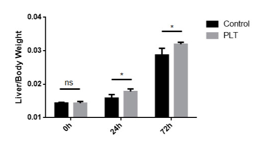
*Results: The liver-to-body weight ratio was significantly higher 24 h and 72 h post-LT in the PLT group compared with the control group (Figure 1). The serum AST and ALT tended to decrease 72h post-LT in the PLT group. The gene expression of cyclin D was significantly increased 72h post-LT in the PLT group, and TNF-a, HGF and IL-6 were increased in the PLT group. Ki-67 positive cells tended to increase in the PLT group 24h and 72h post-LT. There was a significant increase of Mitotic Index in the PLT group 24h post-LT (Figure 2).
*Conclusions: Our results indicate that platelets stimulate liver regeneration after partial LT in rat, and we are to further investigate the mechanism in the future studies.
To cite this abstract in AMA style:
Liang C, Takahashi K, Oda T, Ohkohchi N. Platelets Stimulate Liver Regeneration in a 30% Partial Liver Transplantation Model in Rat [abstract]. Am J Transplant. 2019; 19 (suppl 3). https://atcmeetingabstracts.com/abstract/platelets-stimulate-liver-regeneration-in-a-30-partial-liver-transplantation-model-in-rat/. Accessed February 21, 2026.« Back to 2019 American Transplant Congress


