Multiplexed Imaging Mass Cytometry of the Alloimmune Landscape of Rejection in Clinical Liver Transplantation
J. Emamaullee, N. Ung, C. Man, J. Hoeflich, C. Goldbeck, A. Barbetta, R. Sun, N. Matasci, J. Katz, J. Lee, S. Chopra, L. Sher, S. Asgharzadeh, O. Akbari, Y. Genyk
University of Southern California, Los Angeles, CA
Meeting: 2021 American Transplant Congress
Abstract number: 112
Keywords: Graft-infiltrating lymphocytes, Image analysis, Liver transplantation, Rejection
Topic: Clinical Science » Biomarkers, Immune Assessment and Clinical Outcomes
Session Information
Session Name: Biomarkers, Immune Assessment and Clinical Outcomes - II
Session Type: Rapid Fire Oral Abstract
Date: Sunday, June 6, 2021
Session Time: 4:30pm-5:30pm
 Presentation Time: 4:35pm-4:40pm
Location: Virtual
*Purpose: Rejection continues to be an important cause of graft loss and failure post-liver transplant (LT). Imaging Mass Cytometry (IMC) is an emerging preclinical technique that allows for highly multiplexed simultaneous analysis of immune phenotypes within fixed tissue specimens. In this study, we developed a liver-specific IMC panel as well as an informatics-based analysis pipeline to deeply analyze the immune landscape of chronic rejection (CR) post-LT.
*Methods: A retrospective review of our center identified cases of re-transplant for CR. Regions of interest were identified by our clinical pathologist. A panel of heavy metal-tagged antibodies (Collagen, CD45RA, CD45, CD20, CD3, CD8, CD66a, CD68, CD235, HLA-DR) and a nuclear tag (Iridium) were used simultaneously to label fixed tissue sections for spatial characterization using Hyperion CyTOF IMC. A post-IMC Analysis Pipeline was developed for segmentation and spatial molecular mapping via quantification and co-localization of labeled markers. A total of 18 CR and 5 normal liver (NL) cases were examined.
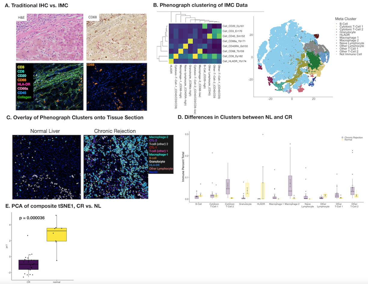
*Results: Examples of IMC staining versus traditional immunohistochemistry (IHC) are shown in Fig. 1A. Detailed quantification of 109,245 cells was completed, of which 30,646 were some type of immune cells after identification. Single cell segmentation followed by Phenograph clustering was used to identify immune subpopulations (Fig. 1B). These clusters were then applied back to the tissue sections to understand density and spatial relationships for each immune population (1C) and quantified (1D), showing significant differences in the proportion of each population between CR and NL (p<0.0001 for each cluster). The entire population of patients was examined using tSNE dimensionality reduction techniques across the IMC marker panel. Modeling via principal component analysis revealed highly consistent immune phenotypes across patients with CR when compared to NL, despite the inherent heterogeneity of clinical LT patients (1E, p=0.000036).
*Conclusions: This study highlights the power of IMC to provide high-resolution, multiplexed analysis of the intragraft immune microenvironment during clinical rejection episodes. Hepatic macrophages have not typically been considered to be involved in CR post-LT, and these data suggest that they may represent a new therapeutic target to treat CR. Further development of IMC has the potential to generate predictive models of clinical outcomes using tissue-specific immune mapping.
To cite this abstract in AMA style:
Emamaullee J, Ung N, Man C, Hoeflich J, Goldbeck C, Barbetta A, Sun R, Matasci N, Katz J, Lee J, Chopra S, Sher L, Asgharzadeh S, Akbari O, Genyk Y. Multiplexed Imaging Mass Cytometry of the Alloimmune Landscape of Rejection in Clinical Liver Transplantation [abstract]. Am J Transplant. 2021; 21 (suppl 3). https://atcmeetingabstracts.com/abstract/multiplexed-imaging-mass-cytometry-of-the-alloimmune-landscape-of-rejection-in-clinical-liver-transplantation/. Accessed February 15, 2026.« Back to 2021 American Transplant Congress

