Indocyanine Green Fluorescence Imaging Allows Quantification of Ischemic Injury During Porcine Normothermic Ex Situ Perfusion
Ajmera Transplant Program, Department of Surgery, Toronto General Hospital, Toronto, ON, Canada
Meeting: 2021 American Transplant Congress
Abstract number: 598
Keywords: Donors, marginal, Graft function, Image analysis, Liver preservation
Topic: Basic Science » Ischemia Reperfusion & Organ Rehabilitation
Session Information
Session Name: Ischemia Reperfusion & Organ Rehabilitation
Session Type: Poster Abstract
Session Date & Time: None. Available on demand.
Location: Virtual
*Purpose: Normothermic Ex Situ Liver Perfusion (NEsLP) is a novel preservation technique for the storage and assessment of grafts retrieved after Donation after Cardio-circulatory Death (DCD). Reliable parameters reflecting graft injury, graft function, and vascular injury during NEsLP are currently missing. Indocyanine Green (ICG) emits fluorescence with near-infrared light and is exclusively cleared by hepatocytes and excreted into the bile without biotransformation. We performed ICG imaging during NEsLP and quantified ICG enhancing and clearance as markers of ischemic liver injury.
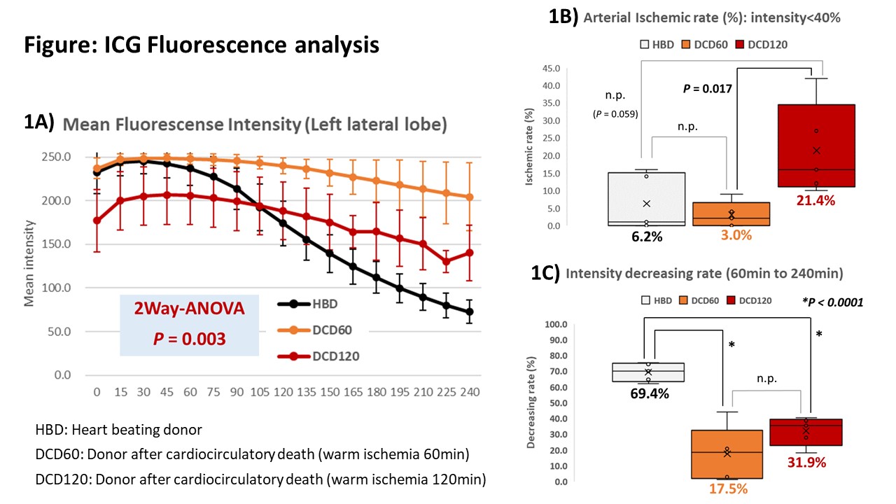
*Methods: 30 to 35 kg male Yorkshire pigs were allocated to 3 groups; Heart beating donor (HBD), DCD 60minutes and DCD 120minutes, each n=5. Following induction of warm ischemia with heparinization, procurement and 2-hour cold storage, the livers were connected to the NEsLP circuit and ICG was injected via the hepatic artery. ICG enhancement and clearance was measured with the SPY Elite® System (Stryker) and analyzed by Image J (National Institutes of Health) software.
*Results: ICG intensity analysis clearly classified three liver patterns (Figure 1A). HBD and DCD60 livers showed rapid intensity increase. Fast clearance of ICG occurred in HBD but not DCD60 livers. DCD120 demonstrated slow enhancement and slow clearance. The arterial ischemic areas were significantly larger in the DCD120 group compared to DCD60 and HBD livers (Figure 1B). ICG excretion rate was significant higher in HBD (69.4%) vs DCD60 (17.5%) vs DCD120 (31.9%) grafts, each P < 0.0001) as shown in Figure 1C.
*Conclusions: ICG imaging during NEsLP is a novel and non-invasive method for classification of DCD liver grafts, which reflects the impairment of arterial perfusion and the metabolic function in the whole graft.
To cite this abstract in AMA style:
Goto T, Noguchi Y, Ganesh S, Mazilescu L, Reichman T, Selzner N, Selzner M. Indocyanine Green Fluorescence Imaging Allows Quantification of Ischemic Injury During Porcine Normothermic Ex Situ Perfusion [abstract]. Am J Transplant. 2021; 21 (suppl 3). https://atcmeetingabstracts.com/abstract/indocyanine-green-fluorescence-imaging-allows-quantification-of-ischemic-injury-during-porcine-normothermic-ex-situ-perfusion/. Accessed February 18, 2026.« Back to 2021 American Transplant Congress

