Imaging OX40+Alloreactive T Cells Provides Early Warning of Organ Transplant Rejection
1Department of Urology, Tokyo Women's Medical University, Tokyo, Japan, 2Department of Radiology, Stanford University, Stanford, CA, 3Department of Otolaryngology-Head and Neck Surgery, Stanford University, Stanford, CA, 4Division of Cardiovascular Medicine, Stanford University, Stanford, CA, 5Division of Blood and Marrow Transplantation, Stanford University, Stanford, CA
Meeting: 2020 American Transplant Congress
Abstract number: 216
Keywords: FK506, Image analysis, Monitoring, T cell graft infiltration
Session Information
Session Name: Biomarker Discovery and Immune Modulation
Session Type: Oral Abstract Session
Date: Saturday, May 30, 2020
Session Time: 3:15pm-4:45pm
 Presentation Time: 3:51pm-4:03pm
Location: Virtual
*Purpose: Diagnosis of organ transplant rejection relies upon biopsy approaches to confirm alloreactive T cell infiltration in the graft. Immuno positron emission tomography (immunoPET) utilizing antibodies conjugated to radioisotopes has the potential to improve early and accurate detection of graft rejection, since it is capable of non-invasively visualizing the dynamic distribution of specific immune markers in the entire body over time. In this work, we identify and characterize the immune checkpoint cell surface molecule OX40 as a surrogate biomarker for alloreactive T cells in organ transplant rejection and monitor its expression utilizing immunoPET.
*Methods: Luciferase+syngeneic and allogeneic hearts were engrafted in bilateral ear pinna on recipients. Graft viability at early time points was assessed with bioluminescent imaging (BLI), and graft infiltrating T cells were characterized by flow cytometry. 89Zr labeled anti-OX40 or IgG1 isotype control antibodies were injected (~50μCi) on d4 after transplantation, and images were captured on d6, d9, and d12. Tacrolimus (Fk) was administered to monitor the subject’s immunosuppression level.
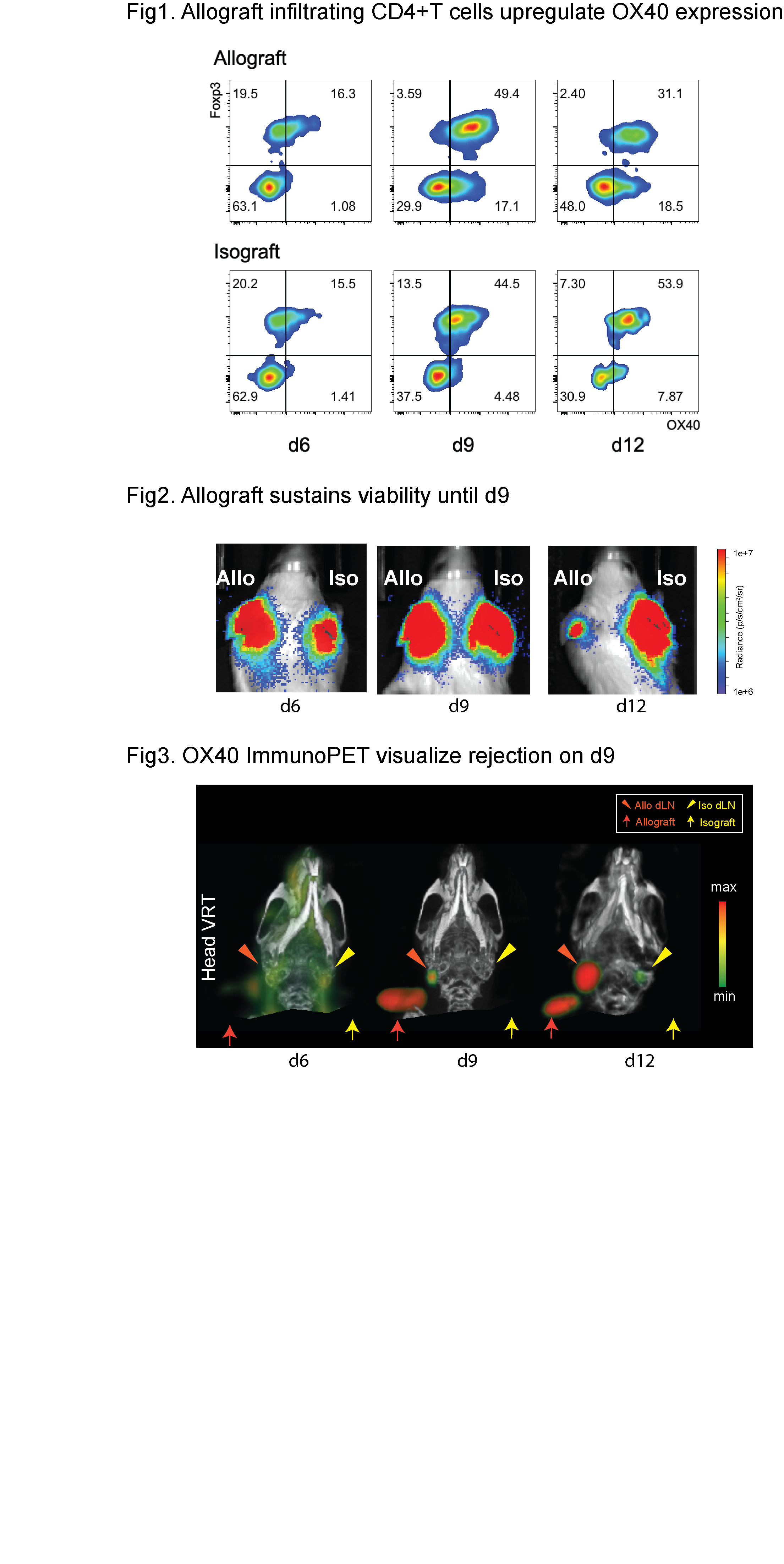
*Results: Flow cytometry showed OX40 upregulation on allograft infiltrating T cells (Fig1). OX40 immunoPET depicted alloreactive T cells in the allograft and allo-draining lymph nodes (dLN), that were not observed in their respective isograft counter parts (n = 7, d9: allograft 8.5±0.8 %ID/g vs isograft 3.4±0.4 %ID/g, p<0.0001, allo dLN 7.9±1.4 %ID/g vs iso dLN 4.1±0.3 %ID/g, p<0.05). The OX40 immunoPET signal identified rejection during the subclinical phase in which rejection was undetectable by either clinical observation or by BLI (Fig2&3). Fk injection significantly reduced tracer uptake in the allograft (n = 5, control 10.6±1.1 %ID/g vs Fk 5.2±1.0 %ID/g, p<0.01). Nonspecific antibody tracer was not able to distinguish rejection, implying high specificity for the target marker.
*Conclusions: This study demonstrates that OX40 immunoPET is a promising approach that may bridge molecular monitoring and morphological assessment for improved transplant rejection diagnosis and monitoring drug dose control.
To cite this abstract in AMA style:
Hirai T, Mayer A, Nobashi TW, Xiao Z, Udagawa T, Seo K, Simonetta F, Baker J, Cheng A, Gambhir SS, Negrin RS. Imaging OX40+Alloreactive T Cells Provides Early Warning of Organ Transplant Rejection [abstract]. Am J Transplant. 2020; 20 (suppl 3). https://atcmeetingabstracts.com/abstract/imaging-ox40alloreactive-t-cells-provides-early-warning-of-organ-transplant-rejection/. Accessed February 13, 2026.« Back to 2020 American Transplant Congress

