Evaluation of UW-Based Solutions Formulated to Be Anti-Apoptotic and Neuroprotective of Retinal Ganglion Cells (RGCs) Following Whole Eye Transplantation (WET) in a Rat Model
1Department of Surgery, Division of Plastic and Reconstructive Surgery, University of Colorado, Anschutz Medical Campus, Aurora, CO, 2Department of Medicine, University of Colorado, Anschutz Medical Campus, Aurora, CO, 3Department of Ophthalmology and Visual Sciences, University of Wisconsin, Madison, WI
Meeting: 2022 American Transplant Congress
Abstract number: 955
Keywords: Nerve allografts, Preservation, Rat, Surgery
Topic: Basic & Clinical Science » Basic & Clinical Science » 20 - VCA
Session Information
Session Time: 7:00pm-8:00pm
 Presentation Time: 7:00pm-8:00pm
Location: Hynes Halls C & D
*Purpose: As vascularized composite allotransplantation (VCA) of an intact optical system to restore vision requires overcoming RGC apoptosis caused by ischemia reperfusion injury, UW and modified UW (mUW: UW + BaCl2 + valproic acid) were assessed as RGC preservation solutions in an orthotopic WET two days post operatively (POD2).
*Methods: Untreated naïve eyes from 14-16-week-old male Brown Norway rats (n=11) were harvested and RGC nuclei were co-stained by immunohistochemistry (IHC) for Brn3a to quantify RGC preservation and phospho-cJun (PNJ) for apoptotic signaling. Prior to donor tissue harvest, donor eyes were intravitreally injected with 10 μL of heparinized saline (n=2), UW (n=3), or mUW (n=3) and transplanted with hemifacial flaps into recipients. At POD2, transplanted and native (recipient, untreated) eyes were enucleated and retinas were prepared for IHC analysis. Continuous variables are presented as mean (± standard deviation) and compared using one-way ANOVA and two sample Z tests.
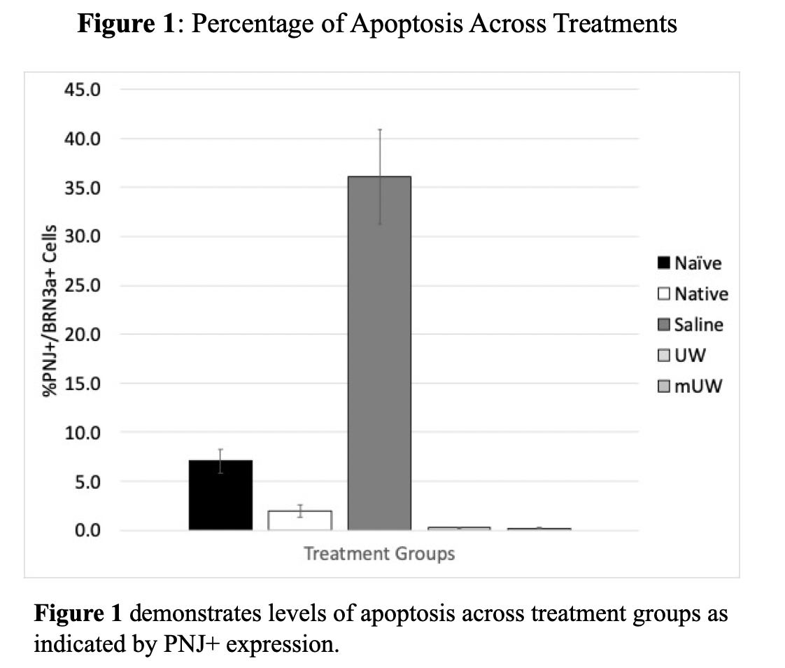
*Results: Naïve retinas expressed 101.8±4.4k BRN3+ RGCs with a 7.1% background expression of PNJ+ signal. At POD2, native recipient retinas show 104.5±5.1k Brn3a+ RGCs with 2% co-staining for PNJ+. In the saline condition, retinas show 63.0±14.4k Brn3a+ RGCs, with 36.1% co-staining for PNJ+. In the UW condition, retinas show 76.2±5.3k Brn3a+ RGCs, with 0.2% co-staining for PNJ+. In the mUW condition, retinas show 75.4±6.4k Brn3a+ RGCs, with 0.2% co-staining for PNJ+.
*Conclusions: We created a novel system to assess RGC preservation and apoptosis post-WET. Injecting UW-based preservation solutions represents a promising strategy for RGC survival following WET. Retinas injected with either mUW and UW solutions showed equal reduction of PNJ+ signaling at POD2 as compared to those injected with heparinized saline (p<0.01). Additionally, RGC survival of eyes injected with UW or mUW showed equal preservation as compared to heparinized saline (p=0.33). Future research will include monitoring Brn3+ and PNJ+ expression at timepoints beyond POD2 as well as the analyzing other apoptotic markers (e.g. activated Caspase-3 and Bcl-2).
To cite this abstract in AMA style:
Khatter NJ, Wang Y, Li B, Owens CR, Jani AH, Huang CA, Nickells RW, Su AA, Washington KM. Evaluation of UW-Based Solutions Formulated to Be Anti-Apoptotic and Neuroprotective of Retinal Ganglion Cells (RGCs) Following Whole Eye Transplantation (WET) in a Rat Model [abstract]. Am J Transplant. 2022; 22 (suppl 3). https://atcmeetingabstracts.com/abstract/evaluation-of-uw-based-solutions-formulated-to-be-anti-apoptotic-and-neuroprotective-of-retinal-ganglion-cells-rgcs-following-whole-eye-transplantation-wet-in-a-rat-model/. Accessed February 26, 2026.« Back to 2022 American Transplant Congress

