Cardiomyocyte Cross-Sectional Area Increases After Orthotopic Cardiac Xenotransplantation and is Not Likely a Significant Contributor to Xenograft Growth up to 9 Months
1Surgery, University of Maryland School of Medicine, Baltimore, MD, 2Department of Medicine, Division of Cardiology, University of Maryland School of Medicine, Baltimore, MD, 3Revivicor, Inc., Blacksburg, VA
Meeting: 2022 American Transplant Congress
Abstract number: 1529
Keywords: Graft survival, Heart/lung transplantation, Post-transplant hypertension
Topic: Basic Science » Basic Science » 13 - Xenotransplantation
Session Information
Session Time: 7:00pm-8:00pm
 Presentation Time: 7:00pm-8:00pm
Location: Hynes Halls C & D
*Purpose: Genetically engineered (GE) pigs are thought to be an alternative organ source for patients in end-stage heart failure unable to receive a timely allograft. However, cardiac xenografts exhibit life-limiting diastolic failure within a short period without addressing potential causes of xenograft growth. Questions remain regarding whether this phenomenon is caused by individual cardiomyocyte hypertrophy.
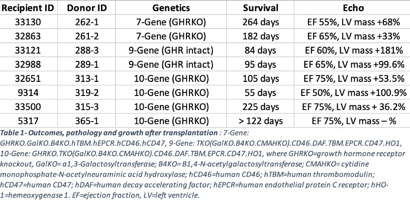
*Methods: GE pig hearts were transplanted orthotopically into weight-matched baboons between 15-30kg, utilizing continuous perfusion preservation prior to implantation (n=8). Neither Temsirolimus nor afterload reducing agents were administered to control for potential causes of post-transplantation growth. GE xenografts contained multiple knockouts ± knockout (KO) for growth hormone receptor (GHR), to address intrinsic xenograft growth and human transgene expression (table 1). TTEs were obtained longitudinally to quantify post-transplantation xenograft growth. Cardiomyocyte cross-sectional area was tabulated and compared to non-transplanted, age-matched controls to measure cellular hypertrophy and compared as ratios (cardiomyocyte transplanted xenograft:non-transplanted control).
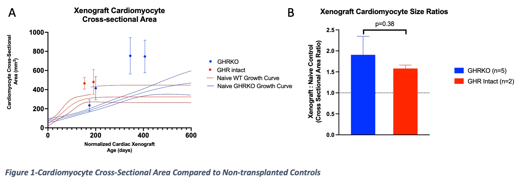
*Results: GHRKO xenografts had a reduced LV mass % increase compared GHR intact xenografts (58.4% ± 27.6 vs. 140.7% ± 58.1, respectively, p=0.041) up to 9 months after transplantation. All cardiomyocytes increased after transplantation, compared to age-matched controls (figure 1a). There was no difference in cardiomyocyte cross-sectional area ratios between GHRKO and GHR intact xenografts (1.9 ± 0.4 and 1.6 ± 0.1, respectively (p=0.38)) (figure 1b).
*Conclusions: These data suggest that individual cardiomyocyte cross-sectional area increases after xenotransplantation regardless of GHR status and does not seem to largely contribute to xenograft growth. Other factors should be considered as more dominant etiologies.
To cite this abstract in AMA style:
Goerlich CE, Griffith B, Treffalls J, Hanna P, Hong SN, Singh AK, Ayares D, Mohiuddin M. Cardiomyocyte Cross-Sectional Area Increases After Orthotopic Cardiac Xenotransplantation and is Not Likely a Significant Contributor to Xenograft Growth up to 9 Months [abstract]. Am J Transplant. 2022; 22 (suppl 3). https://atcmeetingabstracts.com/abstract/cardiomyocyte-cross-sectional-area-increases-after-orthotopic-cardiac-xenotransplantation-and-is-not-likely-a-significant-contributor-to-xenograft-growth-up-to-9-months/. Accessed February 18, 2026.« Back to 2022 American Transplant Congress


