Automatic Quantification of Interstitial Fibrosis in Renal Allograft
1Transplant Research Center, Department of Surgery, Emory University, Atlanta, GA, 2Computer Science Department, Emory University, Atlanta, GA, 3Pathology Department, Northwestern University, Evanston, IL, 4Pathology Department, Emory University, Atlanta, GA
Meeting: 2020 American Transplant Congress
Abstract number: A-305
Session Information
Session Name: Poster Session A: Biomarkers, Immune Assessment and Clinical Outcomes
Session Type: Poster Session
Date: Saturday, May 30, 2020
Session Time: 3:15pm-4:00pm
 Presentation Time: 3:30pm-4:00pm
Location: Virtual
*Purpose: Renal allografts are typically assessed by pathologists using the Banff classification with known interobserver variability. We previously demonstrated the utility of image analysis in assessing interstitial fibrosis; however, improved pipelines and algorithms are needed for routine digital pathology image analysis.
*Methods: Scanned whole slide images were assembled into an image bank using Digital Slide Archive/HistomicsTK (HTK), a system developed by members of our group for image storage, organization, annotation, color normalization, and batch analysis. Banff scores were retrieved from our data warehouse. Fibrosis analysis was conducted on trichrome-stained cortex bounded by glomeruli using HTK batch analysis and compared to the previously used commercial positive pixel count (PPC) algorithm (Aperio). Pearson correlation coefficient and weighted Kappa scores were used to assess agreement between the various methods.
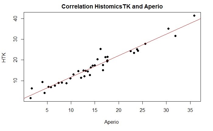
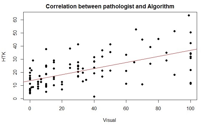
*Results: 103 biopsies with fibrosis spectrum (Banff ci0 n=28, ci1 n=25, ci2 n=25, ci3 n=25) were selected. A moderate agreement between pathologists was observed (inter-pathologist correlation r=0.78 [0.66; 0.86], intra-pathologist correlation r=0.85 [0.66; 0.93]). For Banff ci score, inter-pathologist κ=0.77 [0.70; 0.85] and intra-pathologist κ=0.81 [0.60; 1.00]. Fibrosis analysis by HTK batch analysis was strongly correlated with the PPC algorithm (r=0.97 [0.95; 0.99]) and moderately associated with pathologist assessment (r=0.63 [0.49; 0.73]). Only weak correlations were observed between glomerular filtration rate and pathologist assessment, PPC analysis, and HTK batch analysis.
*Conclusions: We developed a data analysis pipeline allowing rapid automatic fibrosis quantification. Improvement of our quantification algorithm beyond PPC by including more advance artificial intelligence methods is underway to improve correlation with clinical outcome and allow its use to enhanced patient care.
To cite this abstract in AMA style:
Hogan J, Vizcarra J, Tageldin MA, Cooper LA, Gutman DA, Farris AB. Automatic Quantification of Interstitial Fibrosis in Renal Allograft [abstract]. Am J Transplant. 2020; 20 (suppl 3). https://atcmeetingabstracts.com/abstract/automatic-quantification-of-interstitial-fibrosis-in-renal-allograft/. Accessed February 27, 2026.« Back to 2020 American Transplant Congress


