Association of Donor-Derived Cell-Free DNA with Severity of Interstitial Fibrosis and Cortical Atrophy Lesions Following Kidney Transplantation
1University of Minnesota, Minneapolis, MN, 2University of Colorado, Aurora, CO, 3Tampa General Hospital, Tampa, FL, 4Washington University in St. Louis, St. Louis, MO, 5University of Maryland School of Medicine, Baltimore, MD, 6Virginia Commonwealth University, Richmond, VA, 7CareDx, Brisbane, CA, 8University of Texas McGovern Medical School, Houston, TX, 9Intermountain Medical Center, Murray, UT
Meeting: 2021 American Transplant Congress
Abstract number: 1062
Keywords: Histology, Kidney transplantation, Non-invasive diagnosis
Topic: Clinical Science » Kidney » Kidney Complications: Immune Mediated Late Graft Failure
Session Information
Session Name: Kidney Complications: Immune Mediated Late Graft Failure
Session Type: Poster Abstract
Session Date & Time: None. Available on demand.
Location: Virtual
*Purpose: The most common cause of kidney transplant (KT) failure is the poorly characterized histopathologic entity “interstitial fibrosis and tubular atrophy” (IFTA). There are no known unifying mechanisms, nor effective therapy, with diagnosis dependent on biopsy. Incidence of IFTA is ~50% at 1-yr post-KT, 70% at 2-yr, and nearly universal after 10-yr. The combination of IFTA with inflammation is associated with worsened prognosis than fibrosis alone. Considering the multifactorial pathogenesis of IFTA, the aggregation of mechanisms of injury using dd-cfDNA may allow surrogate quantification of inflammation.
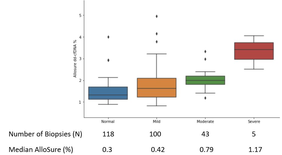
*Methods: 299 patients with paired donor-derived cell-free DNA (dd-cfDNA, AlloSure®; CareDx.) and biopsy (Bx) visits were examined from the Assessing dd-cfDNA monitoring insights of renal allograft with longitudinal surveillance (ADMIRAL study; clinicaltrials.gov: NCT04566055219). Bx performed ≤ 20 days after the dd-cfDNA level, combination of “protocol” and clinical” for cause”. Pathology reports were centrally read and scored, and correlated with histology lesion and dd-cfDNA results. High dd-cfDNA was defined as >0.5% based on previous injury analysis based on ADMIRAL cohort. Interstitial fibrosis (ci) scores were grouped based on the Banff 2018 update. Ci(0)—Interstitial fibrosis in ≤5% of cortical area (normal), ci(1)—Interstitial fibrosis in 6-25% (mild), ci(2)—Interstitial fibrosis in 26-50% (moderate), ci(3)—Interstitial fibrosis in >50% of cortical area (severe).
*Results: dd-cfDNA was significantly elevated when Bx normal patients were compared to those mild to severe (ci). Mild: p= 0.006, moderate: p= 0.01, severe: p= 0.001
*Conclusions: The Banff Classification does not provide precise definition for individual areas of interstitial fibrosis. These results demonstrate a positive correlation between severity of IFTA and allograft injury assessed using dd-cfDNA. Consideration of dd-cfDNA levels with interpretation of histology, may be a useful adjunct in the assessment of interstitial fibrosis, as well as metrics to measure effectiveness of potential therapies.
To cite this abstract in AMA style:
Bu L, Stites E, Bowers V, Alhamad T, Bromberg JS, Gupta G, Moinuddin I, Ghosh S, Tian W, Pai A, Anand S. Association of Donor-Derived Cell-Free DNA with Severity of Interstitial Fibrosis and Cortical Atrophy Lesions Following Kidney Transplantation [abstract]. Am J Transplant. 2021; 21 (suppl 3). https://atcmeetingabstracts.com/abstract/association-of-donor-derived-cell-free-dna-with-severity-of-interstitial-fibrosis-and-cortical-atrophy-lesions-following-kidney-transplantation/. Accessed February 10, 2026.« Back to 2021 American Transplant Congress

