Smartphone-Microfluidic Fluorescence Imaging System for Studying Islet Physiology
University of Virginia, Charlottesville, VA
Meeting: 2022 American Transplant Congress
Abstract number: 1520
Keywords: Image analysis, Islets
Topic: Basic Science » Basic Science » 05 - Translational Cellular Therapies: Islet and Stem Cell Transplantation
Session Information
Session Name: Translational Cellular Therapies: Islet and Stem Cell Transplantation
Session Type: Poster Abstract
Date: Tuesday, June 7, 2022
Session Time: 7:00pm-8:00pm
 Presentation Time: 7:00pm-8:00pm
Location: Hynes Halls C & D
*Purpose: The goal of this study is to develop an innovative smartphone-microfluidic fluorescence imaging (µSFI) system for multi-parameter islet function characterization, which could provide quantitative assessment not only for isolated human islets, but also for new emerging cell biologics, such as stem-cell-derived islets, encapsulated islets, or xenogeneic islets.
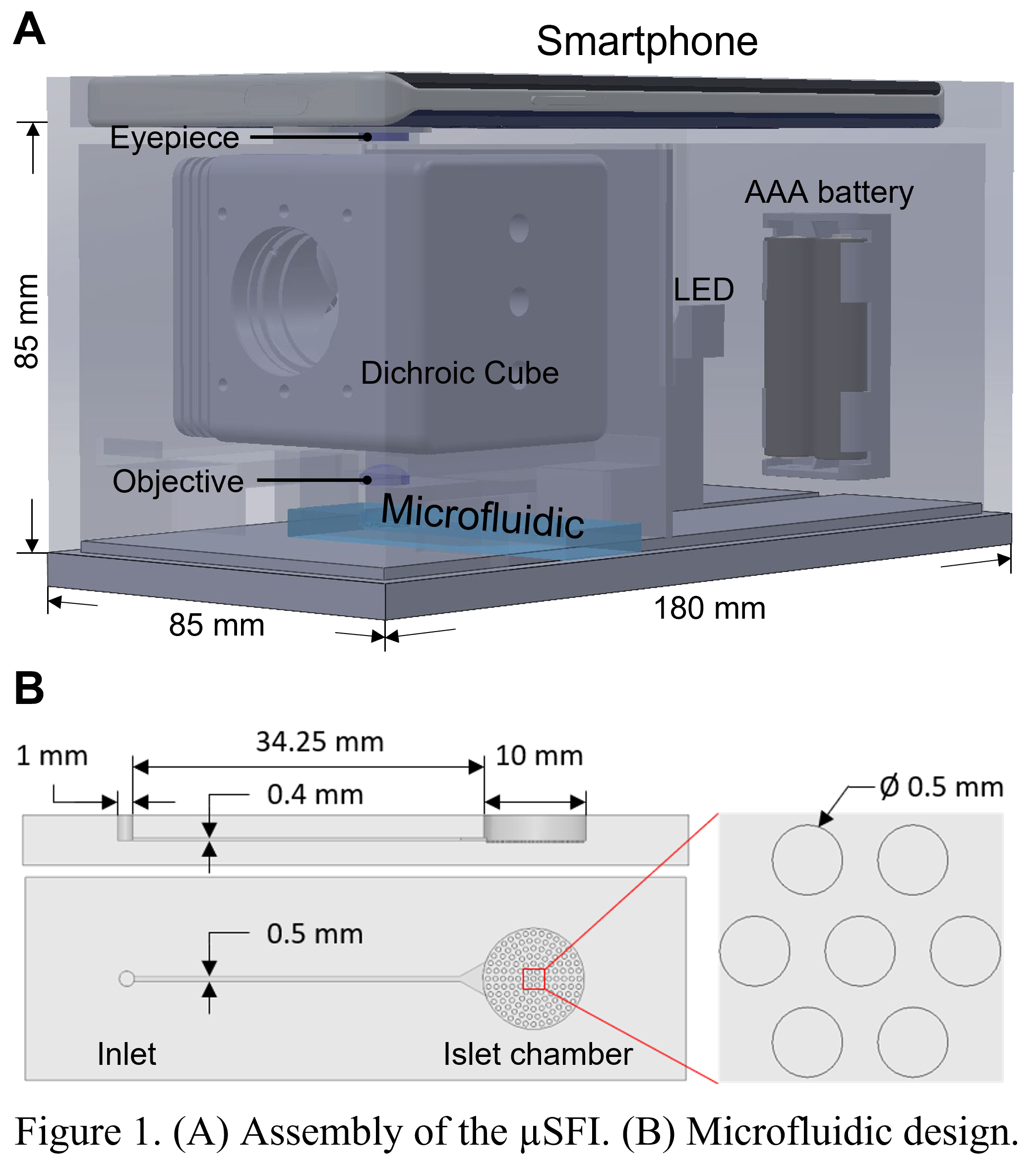
*Methods: The µSFI system consists of a smartphone, two magnification lenses, an LED, and a dichroic cube containing changeable filters. These components are held together by a 3D-printed frame (Fig 1A). Fig 1B shows the design of the microfluidic pumpless biochip for islet imaging. When a drop of liquid (~10 µL) is added to the inlet, due to the surface-tension-induced pressure difference, the fluid will be automatically delivered to the chamber. In addition, the software is developed to control camera/light for image acquisition, analyze signals and generate reports to users. To test the system, we stimulated fluorescence-labeled mouse islets (Fluo-4 indicates [Ca2+]i and Rho123 indicates Ψmito) with high glucose and KCl.
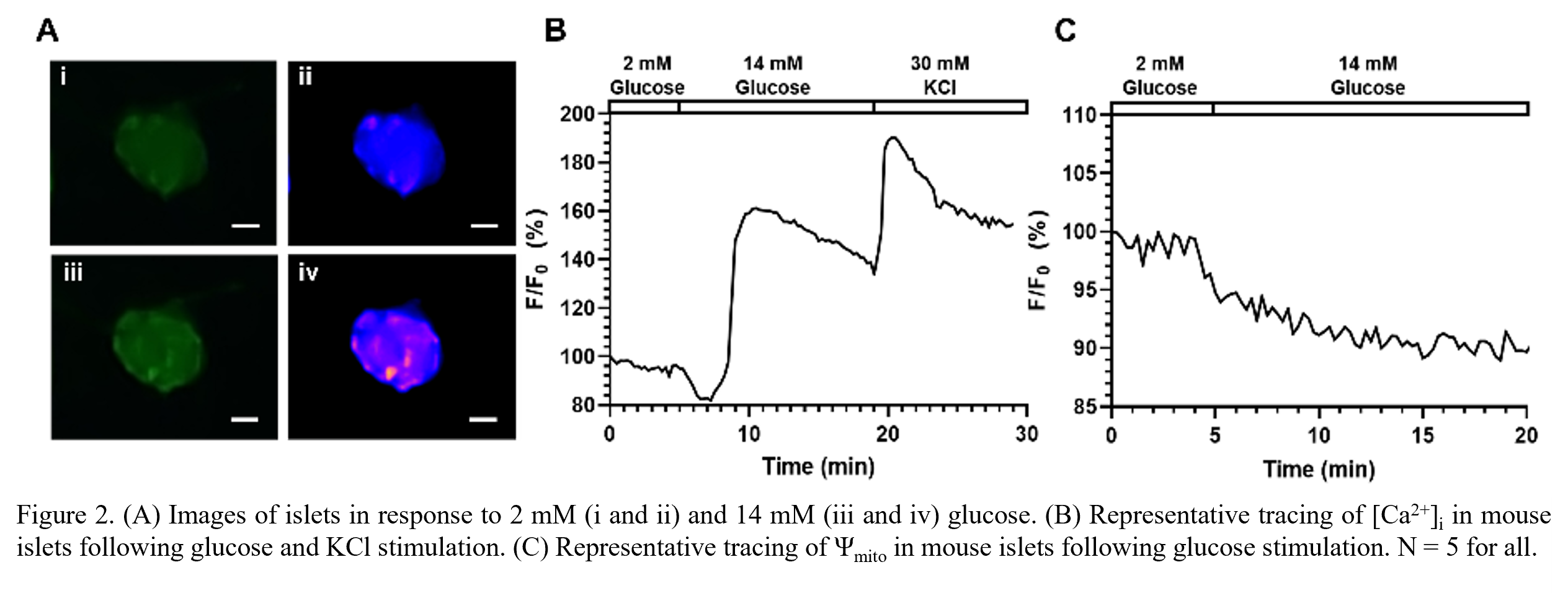
*Results: Images of islets were taken before and after glucose stimulation (Fig 2A), where obvious Fluo-4 signal changes were observed. Under glucose and KCl stimulation (Fig 2B), the curve of Fluo-4 signals showed two obvious peaks. As shown in Fig 2C, Rho123 signals dropped in response to glucose. The results correctly reflected the calcium influx and mitochondrial potential hyperpolarization, which suggested that the system could monitor multi-wavelength fluorescence signals.
*Conclusions: In this study, we introduced the µSFI system to study islet physiology. Compared to conventional fluorescence microscopes, the system presents advantages of low cost, easy-to-operate, and mobility. With all these features, this system would serve as a reliable and user-friendly tool for both on-bench studies of islet physiology and potential clinical applications.
To cite this abstract in AMA style:
Yu X, Xing Y, Zhang Y, Zhang P, He Y, Ramasubramanian MK, Wang Y, Ai H, Oberholzer J. Smartphone-Microfluidic Fluorescence Imaging System for Studying Islet Physiology [abstract]. Am J Transplant. 2022; 22 (suppl 3). https://atcmeetingabstracts.com/abstract/smartphone-microfluidic-fluorescence-imaging-system-for-studying-islet-physiology/. Accessed February 26, 2026.« Back to 2022 American Transplant Congress


