Role of Mitochondrial Transplantation in Enhancing Liver Regeneration Following Partial Hepatectomy
1Transplant Research Institute, James D. Eason Transplant Institute, Department of Surgery, The University of Tennessee Health Science Center, Memphis, TN, 2University of Tennessee/Methodist Transplant Institute, Memphis, TN
Meeting: 2022 American Transplant Congress
Abstract number: 184
Keywords: Ischemia, Machine preservation
Topic: Basic Science » Basic Science » 06 - Tissue Engineering and Regenerative Medicine
Session Information
Session Name: Cellular/Islet Therapies and Tissue Engineering
Session Type: Rapid Fire Oral Abstract
Date: Sunday, June 5, 2022
Session Time: 5:30pm-7:00pm
 Presentation Time: 6:50pm-7:00pm
Location: Hynes Room 310
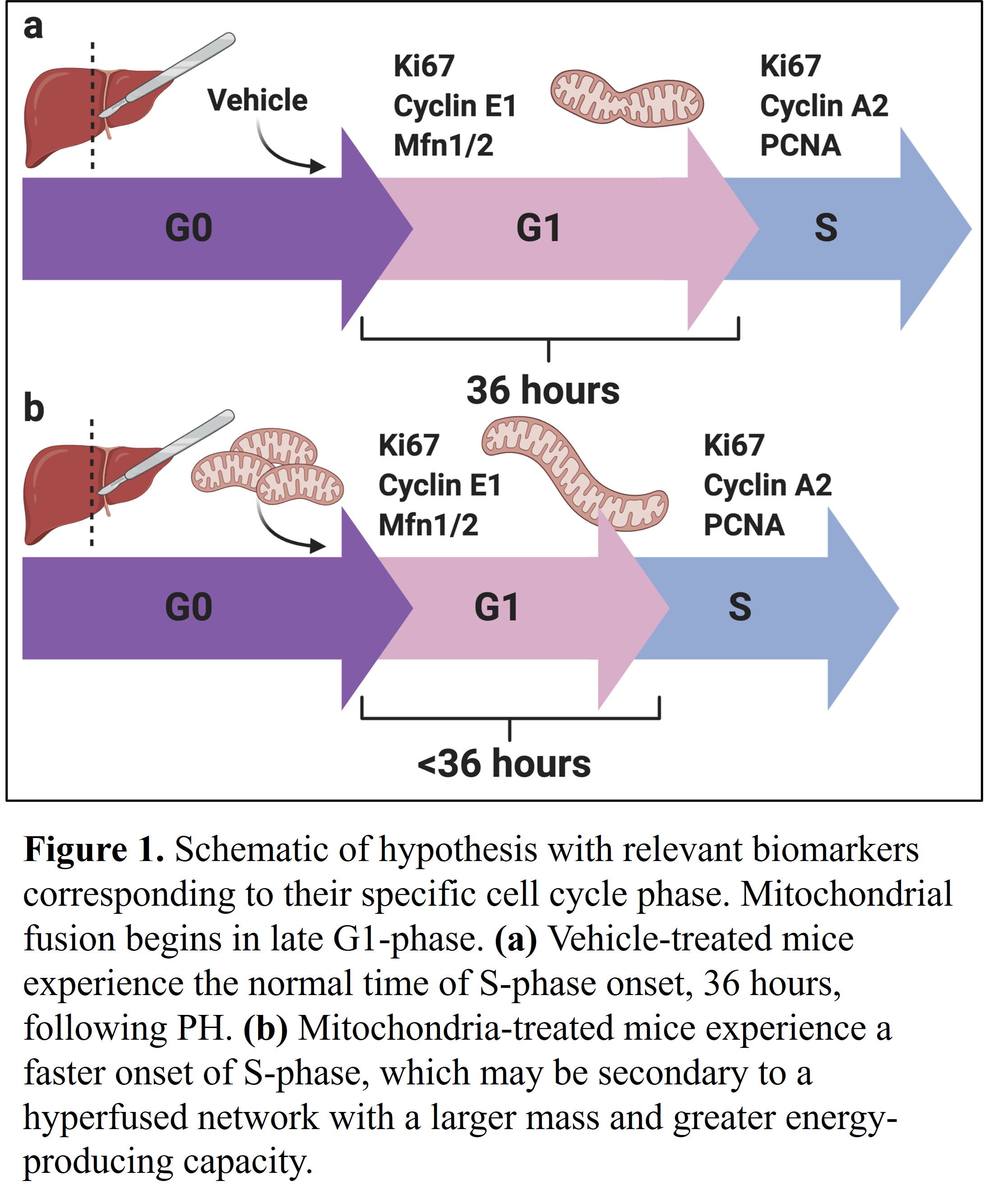
*Purpose: Therapeutic strategies that accelerate the rate of liver regeneration following partial hepatectomy (PH) should be developed to reduce occurrences of small for size syndrome (SFSS), which causes postoperative liver failure that limits the clinical efficacy of surgical modalities for treating liver disease. After PH, as the liver undergoes the regenerative process via cell cycle-driven compensatory hyperplasia, hyperfusion of the hepatocytes’ mitochondrial network is observed in the late G1-phase (Figure1a), which is associated with many functions that promote successful transition into the S-phase. Inhibition of mitochondrial activity attenuates progression through the cell cycle, and mitochondrial dysfunction is often seen in patients that experience postoperative hepatic insufficiency. The project investigated the effects of transplanting exogenous mitochondria on liver regeneration in mice following PH.
*Methods: 8 wk old male C57BL/6 mice underwent partial PH. Mice were divided into two groups vehicle and mitochondria treated. Mice were injected intravenously with mitochondria or 1x PBS one hours after PH. The mice were followed up for either 24 or 48 hrs. Genes for growth markers were measured by RTPCR.
*Results: At 24 hours after PH, mice treated with mitochondria showed higher levels of relative mRNA expression for growth markers Cyclin E1, Cyclin A2, Pcna, and Hif1a compared to vehicle (PBS)-treated mice, suggesting a more robust proliferative response. At 48 hours after PH, the levels of relative mRNA expression for those same markers begin to rise in the vehicle group, while serum albumin levels were higher for the mitochondria-treated mice, indicating improved liver function.
*Conclusions: The data suggests that mitochondrial transplantation can accelerate liver regeneration by inducing a faster onset of the S-phase. The exogenous mitochondria may replace the endogenous mitochondria that were damaged as a result of surgery, provide an excess of energy that allows the remnant hepatocytes to undergo cellular proliferation without having to sacrifice essential homeostatic functions due to limited energy reserves, or fuse with endogenous mitochondria to expedite the formation of the hyperfused mitochondrial network, resulting in a network with a larger mass and greater energy-producing capacity (Figure 1b).
To cite this abstract in AMA style:
Lamanilao G, Watkins C, Kuscu C, Kuscu C, Eason J, Bajwa A. Role of Mitochondrial Transplantation in Enhancing Liver Regeneration Following Partial Hepatectomy [abstract]. Am J Transplant. 2022; 22 (suppl 3). https://atcmeetingabstracts.com/abstract/role-of-mitochondrial-transplantation-in-enhancing-liver-regeneration-following-partial-hepatectomy/. Accessed February 25, 2026.« Back to 2022 American Transplant Congress

