Role of Molecular Microscope in the Interpretation of Endarteritis and Acute Thrombotic Microangiopathy
1Warsaw Medical Univeristy, Warsaw, Poland, 2Gdansk Medical University, Gdansk, Poland, 3ATAGC, University of Alberta, Edmonton AB, AB, Canada, 4Pomeranian Medical University, Szczecin, Poland
Meeting: 2019 American Transplant Congress
Abstract number: C154
Keywords: Biopsy, Gene expression, Kidney transplantation, Rejection
Session Information
Session Name: Poster Session C: Kidney: Acute Cellular Rejection
Session Type: Poster Session
Date: Monday, June 3, 2019
Session Time: 6:00pm-7:00pm
 Presentation Time: 6:00pm-7:00pm
Location: Hall C & D
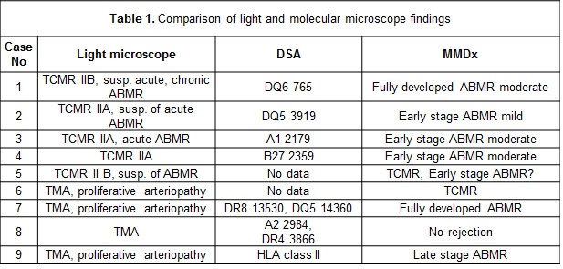
*Purpose: According to the Banff classification, endarteritis (Figure 1A, case 1) may be a manifestation of either: TCMR and ABMR. Acute thrombotic microangiopathy (TMA) constitutes another pattern of injury that potentially may be related to rejection (Figure 1B, case 7). Conventional microscope does not allow for clear identification of these lesions’ background. We assessed molecular phenotypes (MMDx) of v-lesion and TMA in kidney transplant biopsies from the multicenter study “Troubled Polish Kidney Transplants Biopsy”.
*Methods: 90 biopsies ambiguous in the opinion of a clinician or pathologist were included into the study. All light microscope results were reported by one pathologist, molecular phenotype was assessed by Molecular Microscope Diagnostic System (MMDx)
*Results: Among 5 cases of endarteritis (Table 1, case 1-5), one had the molecular phenotype of TCMR, and the other 4 of ABMR. All ABMR cases had positive anti-HLA antibodies: DSA class I in 2 cases, anti DQ in 2 cases (no data from donor typing for DQ). There were 4 TMA cases (Table 1, case 6-9) of which 1 had the molecular phenotype typical of fully-developed ABMR with high titer class II DSA, 1 late stage ABMR without DSA, one of TCMR, and one was not associated with rejection process with moderate titer class I and II DSA. In both groups patients with full developed ABMR lost grafts in 3 month follow up. Case 1 was treated with methylprednisolone (MP) pulses for TCMR and case 7 with MP pulses and 5 plasma exchanges.
*Conclusions: Endarteritis and TMA present serious challenges to treatment selection. The Molecular Microscope is a complementary tool to conventional histology, allowing for more precise lesion interpretation and treatment choice.
To cite this abstract in AMA style:
Ciszek M, Perkowska-Ptasińska A, Deborska-Materkowska D, Debska-Slizien A, Halloran PF, Myslak M. Role of Molecular Microscope in the Interpretation of Endarteritis and Acute Thrombotic Microangiopathy [abstract]. Am J Transplant. 2019; 19 (suppl 3). https://atcmeetingabstracts.com/abstract/role-of-molecular-microscope-in-the-interpretation-of-endarteritis-and-acute-thrombotic-microangiopathy/. Accessed January 1, 2026.« Back to 2019 American Transplant Congress


