Use of Three-Dimensional (3D) Modeling Software for Preoperative Anatomical Assessment in Living Donor Nephrectomy
1University of Rochester School of Medicine and Dentistry, Rochester, NY, 2University of Rochester Medical Center, Rochester, NY
Meeting: 2022 American Transplant Congress
Abstract number: 1390
Keywords: Image analysis, Radiologic assessment
Topic: Clinical Science » Kidney » 41 - Kidney Technical
Session Information
Session Time: 7:00pm-8:00pm
 Presentation Time: 7:00pm-8:00pm
Location: Hynes Halls C & D
*Purpose: Successful and safe completion of living donor nephrectomy depends on donor anatomy. Preoperative imaging is imperative to determine operative feasibility and to avoid unexpected intraoperative findings. The standard of care is cross sectional imaging with Computed Tomography (CT); however this modality may miss fine anatomic details, especially complex venous anatomies. The purpose of this study was to evaluate IRIS (Intuitive Surgical, Sunnyvale CA), adjunctive software to the da Vinci surgical system which provides 3D volumetric reconstructions of renal anatomy. This modality was compared to traditional CT for detecting anatomic variants preoperatively.
*Methods: All institutional living donor nephrectomies, including laparoscopic and robotic assisted, utilizing the IRIS software for the years 2020 and 2021 were reviewed, totaling 36 patients. Two patients did not undergo nephrectomy and were excluded. Charts were reviewed to determine operative laterality. Prior to further chart review IRIS scans of all patients were graded. Anatomy was assessed on the operative side for multiplicity, configuration, and anatomic variants of the renal hilar structure (renal artery, renal vein, and ureter). Preoperative CT reports were reviewed, and the same renal anatomic descriptions were gathered based on radiologist interpretation. Finally, operative reports were reviewed for encountered anatomies and unexpected intraoperative findings.
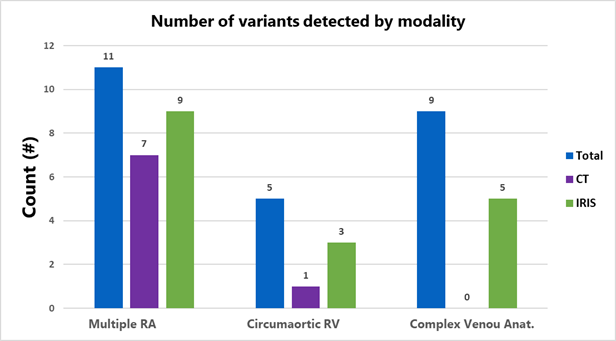
*Results: Operatively encountered variants among the cohort included 11 incidences of multiple renal arteries (RA), 5 of circumaortic renal veins, and 9 of other complex renal venous anatomy, which included multiple or large lumbar veins or suprarenal veins, aneurysmal veins, and non-typical branching of the gonadal vein. In the cohort there were no duplicated renal veins, and all ureteral anatomy was traditional. Across modalities, IRIS detected more total variants than traditional CT. IRIS detected 9/11 (81%) of multiple RA vs 7/11(63%) detection for CT; 3/5(60%) of circumaortic veins vs 1/5(20%); 5/9(55%) of complex venous anatomies vs 0%.
*Conclusions: Our data demonstrates that 3D volumetric reconstructions from traditional CT scans such as constructed by the IRIS software are a promising modality for preoperative anatomic assessment in living donor nephrectomy and may demonstrate superiority for the detection of certain anatomic variants, especially complex venous anatomies.
To cite this abstract in AMA style:
McCabe M, Dokus K, Nair A, Pineda-Solis K, Venniro E, Taylor J, Orloff M, Hernandez R, Kashyap R. Use of Three-Dimensional (3D) Modeling Software for Preoperative Anatomical Assessment in Living Donor Nephrectomy [abstract]. Am J Transplant. 2022; 22 (suppl 3). https://atcmeetingabstracts.com/abstract/use-of-three-dimensional-3d-modeling-software-for-preoperative-anatomical-assessment-in-living-donor-nephrectomy/. Accessed February 27, 2026.« Back to 2022 American Transplant Congress

