Technical Report to Increased Utilization of Injured Kidneys: CT Scan to Evaluate Organ and Reduce Discard
Surgery, Columbia Vagelos College of Physicians and Surgeons, New York, NY
Meeting: 2022 American Transplant Congress
Abstract number: 1400
Keywords: Cadaveric organs, Kidney transplantation, Renal injury, Resource utilization
Topic: Clinical Science » Kidney » 41 - Kidney Technical
Session Information
Session Time: 7:00pm-8:00pm
 Presentation Time: 7:00pm-8:00pm
Location: Hynes Halls C & D
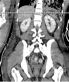
*Purpose: To reduce discard of injured kidneys with parenchymal injury or perinephric hematoma . Young donors with good function but injury are at high risk of discard.Evaluating the CT angiogram of the kidney is a simple and effective tool to fully evaluate these kidneys for major parenchymaI injuries and determine true extent of injury. These organs often accumulate cold time which further increase the chance of discard.
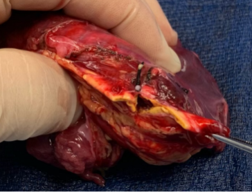
*Methods: Analysis of two donors with traumatic injuries. Donor features such as age, weight, renal function, anatomy, serology, urine production. High refusal led to long cold time and discard risk. Based on these CT images: kidneys were accepted for transplant pending back table prep. The organ function and bleeding parameters are presented estimated blood loss (EBL), cold time and complications associated with use of these organs.
*Results: Donor A: 4-year-old brain dead donor fell from 10th floor balcony. Splenic laceration, 50% parenchymal involvement AAST grade II injury. Laceration of right adrenal gland > 50% and parenchymal laceration 1.4 cm (AAST grade III injury). Donor B: 20-year-old MVC vs dump truck. Extensive TBI/IVH/SDH. Had splenectomy, left renal laceration grade III. Family elected to proceed with DCD. See table. Both kidneys functioned, no complications, no excess bleeding. Blood transfusion for post-operative hematocrit 19.3 lowest without hemodynamic changes in recipient of organ B. All organs showed graft function. Graft A: 5 months of follow up: baseline creat 1.3.
*Conclusions: CT scan images to visualized injured kidney provide additional source of information showing cortical integrity despite perinephric hematoma leading to increase utilization without adversely affecting patient or graft outcomes. The approach demonstrates feasibility in reducing discard of injured high-quality organs.
| Donor | Age (y) | Wt (kg) | Bld Type | Lateral | Import | Dist. | EBL |
| A | 4 | 20 | O | R | No | 2.69 | 100 |
| B | 20 | 80 | O | L | Yes | 667 | 150 |
| Donor | Rec. seq# | LOS | Transfusion | Init. hct | lowest hct post op | CIT | Complications |
| A | 136 | 4 | No | 28.9 | 22.6 | 25h 19 | No |
| B | 3895 | 7 | Yes | 27.3 | 19.3 | 32h 44 | No |
| Rec age(A)
52
|
KDPI (A)
33%
|
||||||
| Rec
age(B)
46
|
KDPI
(B)
5%
|
To cite this abstract in AMA style:
Whittaker VE. Technical Report to Increased Utilization of Injured Kidneys: CT Scan to Evaluate Organ and Reduce Discard [abstract]. Am J Transplant. 2022; 22 (suppl 3). https://atcmeetingabstracts.com/abstract/technical-report-to-increased-utilization-of-injured-kidneys-ct-scan-to-evaluate-organ-and-reduce-discard/. Accessed February 13, 2026.« Back to 2022 American Transplant Congress


