Severe Refractory Skin Eruption Treated With Plasmapheresis in a Multivisceral Transplant Patient
1Miam Transplant Institute, University of Miami Miller School of Medicine, Miami, FL
2Pediatrics, University of Miami Miller School of Medicine, Miami, FL
3Surgery Critical Care, University of Miami Miller School of Medicine, Miami, FL.
Meeting: 2015 American Transplant Congress
Abstract number: A280
Keywords: Graft-versus-host-disease, Multivisceral transplantation, Plasmapheresis
Session Information
Session Name: Poster Session A: Small Bowel All Topics
Session Type: Poster Session
Date: Saturday, May 2, 2015
Session Time: 5:30pm-7:30pm
 Presentation Time: 5:30pm-7:30pm
Location: Exhibit Hall E
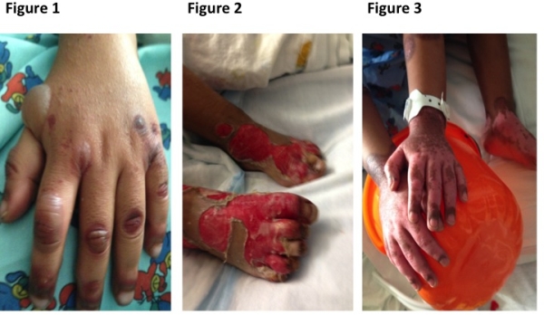
3 year old African American male who underwent a multivisceral transplant due to gastroschisis. Crossmatch for T and B cells, PRA, DSA were negative. Induction was with anti-thymocyte globulin (ATG), rituximab and steroids. Postoperative course was complicated by severe acute rejection. He received 2 doses of Basixilimab and steroid cycle. Given no improvement, 2 more doses of ATG were administered. Mucosal healing was complicated by an enteric stricture in the 3rd portion of the duodenum. He developed bullous lesions starting on dorsal surface of hands and feet bilaterally. Skin biopsy showed necrotic epithelium with superficial perivascular inflammation (lymphocytes, plasma cells and eosinophils). Diagnosis included bullous erythema multiforme (EM)-like drug eruption vs. GVHD. Given chimerism and rectal biopsies were negative for GVHD, drug eruption was favored. IV Zosyn/Tylenol were new medicatons when lesions appeared and suspected. Both were discontinued. Lesions coalesced leaving eschar. Diagnosis broadened to include infection vs. Steven Johnson Syndrome (SJS) vs. nutritional vs. autoimmune process. Bullous impetigo was considered and a course of Clindamycin was given. A trial of IV steroids and high dose IVIG was given for suspected SJS. Vitamin levels were normal. Biopsy done with direct immunofluorescence was normal. Tacrolimus was discontinued for 3 weeks. Lesions progressed to scalp, torso and perineum with epidermolysis leading to 2nd degree burns. Given worsening picture, immunologic phenomenon was suspected. Plasmapheresis/Rituximab (5 cycles) was performed to remove possible offending toxins. Wound care continued to be judiciously performed. Thereafter, skin lesions markedly improved.
Conclusion: Plasmapheresis is an option for treatment of severe skin lesions in transplant patients when other modalities are unsuccessful.
To cite this abstract in AMA style:
Garcia J, Beduschi T, Tekin A, Fan J, Nishida S, Selvaggi G, Ciancio G, Burke G, Katsoufis C, Pizano L, Vianna R. Severe Refractory Skin Eruption Treated With Plasmapheresis in a Multivisceral Transplant Patient [abstract]. Am J Transplant. 2015; 15 (suppl 3). https://atcmeetingabstracts.com/abstract/severe-refractory-skin-eruption-treated-with-plasmapheresis-in-a-multivisceral-transplant-patient/. Accessed February 17, 2026.« Back to 2015 American Transplant Congress
