Renal Fibroblast Activation and Survival by Anti-MHC Class I Antibody: The Role of mTOR Signaling Pathway.
Medicine/Surgery, University of Alabama at Birmingham, Birmingham, AL.
Meeting: 2016 American Transplant Congress
Abstract number: A9
Keywords: Alloantibodies, Fibrosis, Kidney transplantation, MHC class I
Session Information
Session Type: Poster Session
Date: Saturday, June 11, 2016
Session Time: 5:30pm-7:30pm
 Presentation Time: 5:30pm-7:30pm
Location: Halls C&D
Background: Antibody-mediated injury (AMI) is an important contributor to late kidney allograft loss, and is accompanied by interstitial fibrosis (IF) and tubular atrophy (TA). However, a link between anti-donor HLA antibodies and graft fibrosis is not known. We hypothesized that circulating anti-HLA antibodies, in addition to effects on endothelial cells, could activate and differentiate renal fibroblasts, thus contributing to graft fibrosis.
Methods: Primary mouse kidney fibroblasts (mKFB) from Balb/c mice were cultured in the presence of anti-MHC Class I antibody (anti-H2d) or isotype control. Cell proliferation was measured by Brdu incorporation. Gene expression was analyzed by RTPCR. AKT and mTOR activation were detected by western blot, as well as survival molecule Bcl-xl.
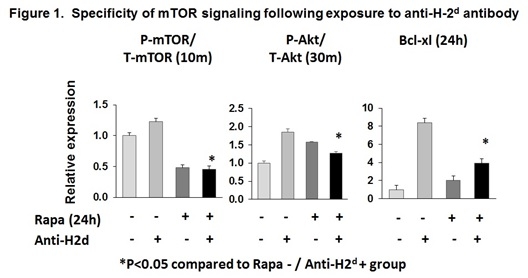
Results: Compared to isotype control, exposure of mKFB to anti-H2dantibody (48h,1[micro]g/ml) significantly induced cell proliferation (1.3±0.04-fold, p=0.01). This was accompanied by marked increases in gene expression of αSMA (3.8±0.2-fold, p=0.01), S100A4 (2.0±0.1-fold, p=0.01), and fibronectin1 (2.0±0.1-fold, p=0.01), indicating myofibroblast activation. mKFB proliferation and activation were associated with dramatic phosphorylation of AKT at ser473 (23.1±2.1-fold; p=0.02) and thr308 (22.4±0.1-fold; p=0.02).This was accompanied by phosphorylation of mTOR (1.2±0.0-fold at 10min, p=0.03) but without significant change in downstream molecules p70S6K and S6RP, suggesting that the antibody-mediated proliferation signal is mediated by other pathways. However, anti-H2dantibody enhanced expression of anti-apoptotic protein Bcl-xl (2.2±0.03-fold, p=0.01 compared to isotype control). Moreover, pretreatment with mTOR inhibitor rapamycin significantly reduced anti-H2dantibody-induced mTOR/AKT phosphorylation and Bcl-xl expression (Figure 1).
Conclusions: Our results demonstrate the critical effect of anti-MHC class I antibody on renal fibroblasts with a potent ability to induce cell proliferation, activation, and survival through the mTOR/AKT signaling pathway. Further understanding of this mechanism may shed light on novel interventions to mitigate allograft fibrosis in the setting of AMI. 
CITATION INFORMATION: Chen J, Zmijewska A, Mannon R. Renal Fibroblast Activation and Survival by Anti-MHC Class I Antibody: The Role of mTOR Signaling Pathway. Am J Transplant. 2016;16 (suppl 3).
To cite this abstract in AMA style:
Chen J, Zmijewska A, Mannon R. Renal Fibroblast Activation and Survival by Anti-MHC Class I Antibody: The Role of mTOR Signaling Pathway. [abstract]. Am J Transplant. 2016; 16 (suppl 3). https://atcmeetingabstracts.com/abstract/renal-fibroblast-activation-and-survival-by-anti-mhc-class-i-antibody-the-role-of-mtor-signaling-pathway/. Accessed February 22, 2026.« Back to 2016 American Transplant Congress
