Non-Invasive Measurement of Transplant Kidney Fibrosis Using Photoacoustic Imaging
1St. Michael's Hospital, Toronto, ON, Canada, 2Ryerson University, Toronto, ON, Canada
Meeting: 2021 American Transplant Congress
Abstract number: 423
Keywords: Allocation, Fibrosis, Kidney, Kidney transplantation
Topic: Clinical Science » Kidney » Kidney Technical
Session Information
Session Name: Kidney Deceased Donor Allocation 2
Session Type: Poster Video Chat
Date: Sunday, June 6, 2021
Session Time: 7:30pm-8:30pm
 Presentation Time: 7:40pm-7:50pm
Location: Virtual
*Purpose: The presence of donor kidney fibrosis adversely impacts transplant outcome. Biopsy remains the only method that can assess fibrosis, despite being subjected to sampling bias as it measures <1% of the kidney volume. In this work we propose to use collagen-specific photoacoustic (PA) imaging as a non-invasive and contrast-free tool for measuring kidney fibrosis. PA imaging relies on the detection of sound waves produced by short laser burst illuminations of the kidneys.
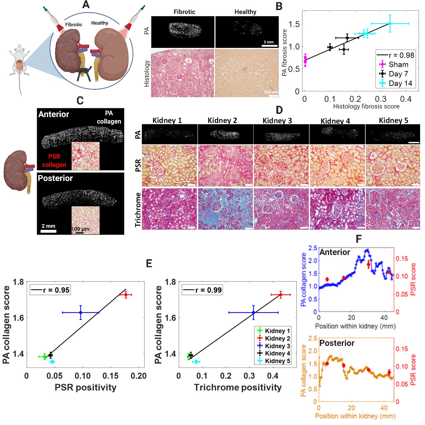
*Methods: This technology was tested in a unilateral ureteral obstruction (UUO) model of mouse kidney fibrosis (Fig. 1A). Surgery was performed on n = 10 mice and the left ureter was obstructed for either 7 or 14 days. Comparisons with sham surgery performed on n = 5 mice were also conducted. The kidneys were procured, and PA imaging was performed before sectioning each kidney for histology. Using multiwavelength PA images acquired from a handheld probe, a novel collagen estimation algorithm was implemented to compute renal fibrotic burden. Its accuracy in estimating collagen was compared to the gold standard histology. The algorithm was further tested in human radical nephrectomy specimens, as well as in whole human kidneys in a setting mimicking the ex vivo phase of kidney transplantation surgery.
*Results: PA-measured fibrosis scores correlated strongly with gold standard histology measurements, being able to detect increasing levels of fibrosis (Fig. 1B). PA imaging of the cortex of whole human kidneys was possible by imaging over the entire kidney surface (Fig. 1C). Our PA technique was able to accurately capture inter-kidney variation in fibrosis levels (Fig. 1D) with excellent accuracy (Fig. 1E). As seen in mouse kidneys, PA imaging could also to detect intra-renal differences in human kidney fibrosis (Fig. 1F).
*Conclusions: This study demonstrates for the first time the potential of PA imaging as a non-invasive renal fibrosis measurement tool. By directly imaging collagen, this technology is non-invasive, contrast-free, sensitive to a wide range of fibrosis burdens and can be performed in just minutes without impeding the transplant procedure. Taken together, these findings suggest that PA imaging may be able to play an important role in assessing the level and spatial distribution of fibrosis in ex vivo donor kidneys. A clinical trial to assess the feasibility of ex vivo donor kidney PA imaging at the time of transplant to assess allograft fibrosis and outcomes is scheduled to commence early next year.
To cite this abstract in AMA style:
Hysi E, He X, Krizova A, Ordon M, Pace KT, Farcas M, Kolios MC, Yuen DA. Non-Invasive Measurement of Transplant Kidney Fibrosis Using Photoacoustic Imaging [abstract]. Am J Transplant. 2021; 21 (suppl 3). https://atcmeetingabstracts.com/abstract/non-invasive-measurement-of-transplant-kidney-fibrosis-using-photoacoustic-imaging/. Accessed February 14, 2026.« Back to 2021 American Transplant Congress

