Changes in Diastolic Function and Cardiac Geometry After Pediatric Kidney Transplant: A Longitudinal Study
1Nephrology, Children's National Hospital, Washington DC, DC, 2Cardiology, Children's National Hospital, Washington DC, DC
Meeting: 2022 American Transplant Congress
Abstract number: 112
Keywords: Echocardiography, Heart, Kidney transplantation, Pediatric
Topic: Clinical Science » Kidney » 43 - Kidney: Pediatrics
Session Information
Session Time: 5:30pm-7:00pm
 Presentation Time: 6:50pm-7:00pm
Location: Hynes Ballroom C
*Purpose: Kidney transplant recipients are at increased risk for cardiovascular disease but systolic function is often preserved and may not reveal cardiac dysfunction until it is severe. Changes in diastolic function and cardiac geometry have not been well studied in pediatric transplant recipients. We conducted a prospective controlled longitudinal study to investigate changes in diastolic function and cardiac geometry after kidney transplant, as compared to pre-transplant and healthy controls.
*Methods: Transplant recipients (3-21 years old) had echocardiograms performed pre-transplant and at 1, 18, 30, and 42 months post-transplant. A single echocardiogram was performed for each healthy control participant. Indicators of diastolic function (E/e’ lateral, E/A) and cardiac geometry (interventricular septal end diastole diameter [IVSd], left ventricular posterior wall end diastole diameter [LVPWd], left atrial dimension [LA] z-scores) were evaluated.
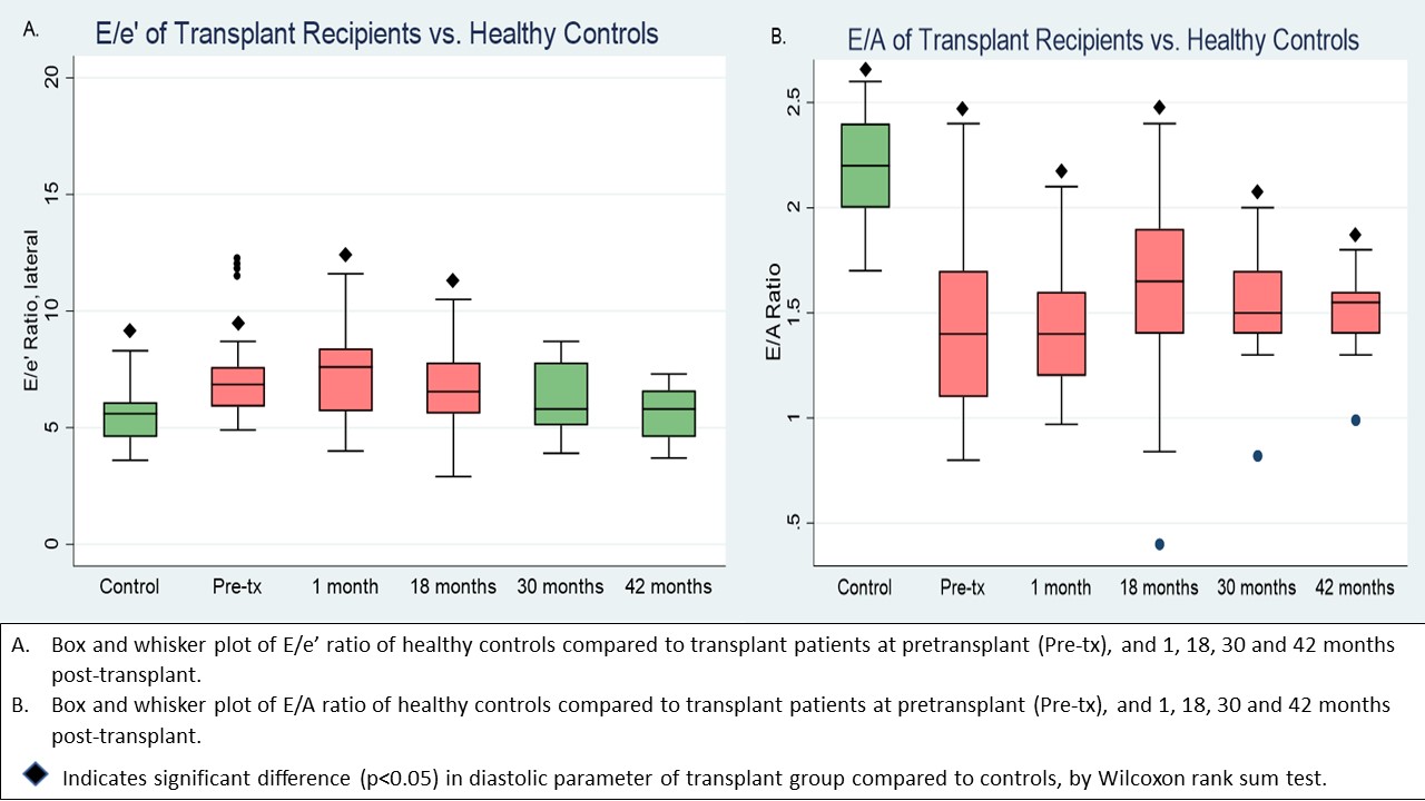
*Results: The study included 41 transplant participants and 26 healthy controls. Of 41 transplant recipients, 15 had received hemodialysis (HD), 13 peritoneal dialysis (PD) and 13 pre-emptive transplant. E/e’ and E/A of patients were worse than controls at pre, 1, and 18 months post-transplant; E/e’ improved by 30 months while E/A remained reduced (Figure). Systolic dysfunction (ejection fraction<55% or fractional shortening<25%) was present in 7.5% pre-transplant, while no patients exhibited systolic dysfunction 1-30 months post-transplant. Exposure to HD was associated with worse E/e’ pre and post-transplant. After transplant, impaired E/e’ was associated with steroid exposure (β 2.0 p=0.01) and abnormal E/A with metabolic syndrome (β -0.14 p=0.03). Cardiac geometry was abnormal (LVPWd, IVSD, and/or LA >95th%) in 43.2% pre-transplant, and remained abnormal in 42.2%, 31.2%, 30.7%, and 30.0% of patients at 1, 18, 30, and 42 months post-transplant. Increased LVPWd was associated with hypertension (β 0.46 p=0.01) and steroid exposure (β 0.91 p=0.02). Enlarged LA was associated with glomerular diagnosis (OR 4.1 p=0.02), steroid exposure (OR 4.2 p=0.04), and left ventricular hypertrophy (OR 3.0 p=0.02).
*Conclusions: Pediatric kidney transplant recipients have worse diastolic function and altered cardiac geometry compared to healthy peers, while systolic function is preserved. Exposure to HD is associated with diastolic changes pre and post-transplant. Attention to early diastolic changes may provide an opportunity to intervene before advanced changes occur.
To cite this abstract in AMA style:
Sgambat K, Clauss S, Moudgil A. Changes in Diastolic Function and Cardiac Geometry After Pediatric Kidney Transplant: A Longitudinal Study [abstract]. Am J Transplant. 2022; 22 (suppl 3). https://atcmeetingabstracts.com/abstract/changes-in-diastolic-function-and-cardiac-geometry-after-pediatric-kidney-transplant-a-longitudinal-study/. Accessed February 10, 2026.« Back to 2022 American Transplant Congress

