Case Series Of Total Laparoscopic-hand Assisted Living Donor Right Hepatectomy: First 19 Cases At A Single Center In Usa
1Surgery, Division of Transplantation, University of Massachusett, Worcester, MA, 2Surgery, Division of Transplantation, University of Massachusetts, Worcester, MA
Meeting: 2019 American Transplant Congress
Abstract number: B353
Keywords: Donation, Laparoscopy, Liver transplantation, Outcome
Session Information
Session Name: Poster Session B: Liver: Living Donors and Partial Grafts
Session Type: Poster Session
Date: Sunday, June 2, 2019
Session Time: 6:00pm-7:00pm
 Presentation Time: 6:00pm-7:00pm
Location: Hall C & D
*Purpose: One of the major limitations of living-donor liver donation is the associated morbidity of the procedure. Although left and left-lateral hepatectomy has been performed by several centers for pediatric liver transplantation, right-laparoscopic liver resection has been successfully done by very few centers. Here we described our first 19 cases of total hand-assisted laparoscopic right hepatectomy for adult liver transplantation.
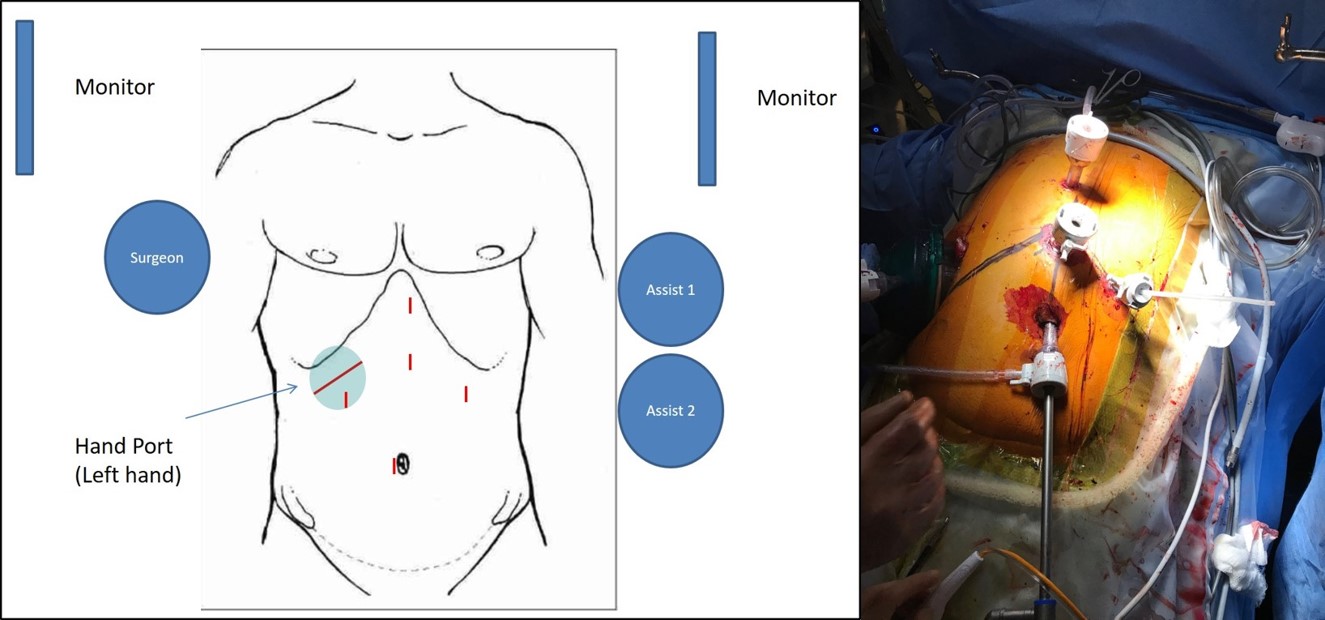
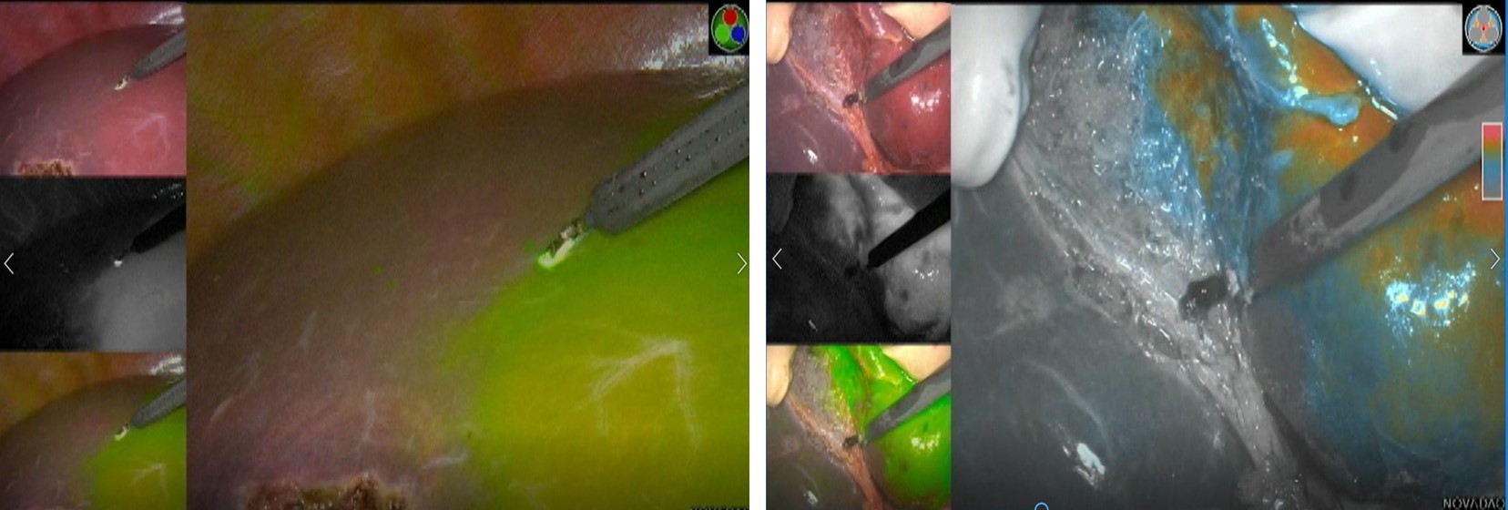
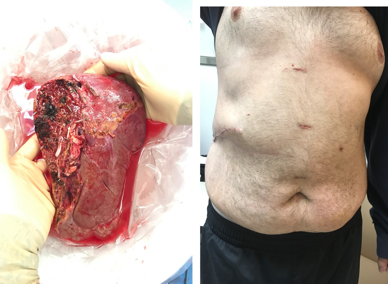
*Methods: We retrospectively reviewed the medical records of all laparoscopic right lobe living donor liver resections performed at UMass Memorial Medical Center between between 10/25/2016 and 11/30/2018. A total of 19 living donors underwent hand-assisted right hepatectomy for adult LDLT. We recorded the biliary anatomy according to the Choi Classification, the portal anatomy according to Cheng classification, length of stay, and post-op complications according to the Clavien-Dindo classification. A hand gelport was placed on the right subcostal incision and four laparoscopic ports were introduced (figure 1). The operations were done laparoscopically until removal of graft. Parenchymal division was performed after delineation of the ischemic line (Cantlie’s) with ICG under visualization with the Spy scope system (figure 2). Division was performed using an ultrasonic dissector (CUSA), bipolar coagulation, clips, and endovascular stapler device. A real-time near infra-red indocyanine green (ICG) fluorescent cholangiography (Pinpoint Spy system ™, Novadaq, Ontario-CA) and an intra-op traditional cholangiography (with C-arm and contrast medium injection) were performed to identify the bifurcation and to cut the right hepatic duct. The bile duct was divided after fluoroscopic ICG-cholangiogram. The right liver graft was removed through the 10-cm right subcostal incision (figure 3).
*Results: The median age of donors was 46 years, median graft vs recipient weight ratio was 1.0. The media estimated blood loss was 500ml (mean 512ml). No blood products were given except in two cases. The postoperative course was uneventful in all cases except two because of bile leak. The donors were discharged at median postoperative day 8. The donor liver function tests returned close to baseline within a week post donation. There was no graft loss and no delayed graft function.
*Conclusions: These cases offer evidence that hand-assisted laparoscopic living donor right hepatectomy can be safely performed. This technique may allow early recovery for the living donor and increase the rate of living donor liver donation.
To cite this abstract in AMA style:
Martins PN, Movahedi B, Litwin D, Bozorgzadeh A. Case Series Of Total Laparoscopic-hand Assisted Living Donor Right Hepatectomy: First 19 Cases At A Single Center In Usa [abstract]. Am J Transplant. 2019; 19 (suppl 3). https://atcmeetingabstracts.com/abstract/case-series-of-total-laparoscopic-hand-assisted-living-donor-right-hepatectomy-first-19-cases-at-a-single-center-in-usa/. Accessed February 10, 2026.« Back to 2019 American Transplant Congress


