Nanoparticle Mediated Drug Delivery for Lung Transplantation
1Surgery, Center for Transplantation Sciences, Massachusetts General Hospital/Harvard Medical School, Boston, MA, 2Medicine, Transplant Research Center, Brigham and Women's Hospital, Boston, MA, 3Surgery, Center for Transplantation Sciences, Division of Cardiac Surgery, Massachusetts General Hospital/Harvard Medical School, Boston, MA
Meeting: 2021 American Transplant Congress
Abstract number: 307
Keywords: Immunosuppression, Lung, Perfusion solutions
Topic: Basic Science » Biomarker Discovery and Immune Modulation
Session Information
Session Name: Biomarkers and Cellular Therapies
Session Type: Rapid Fire Oral Abstract
Date: Tuesday, June 8, 2021
Session Time: 4:30pm-5:30pm
 Presentation Time: 5:00pm-5:05pm
Location: Virtual
*Purpose: Severe primary graft dysfunction (PGD) affects 30% of lung transplant patients. Pulmonary alveolar macrophages play a significant role in lung PGD, but systemic immunotherapy is inadequate in suppressing them. We explored using nanoparticles (NP) as a means of direct allograft immunosuppression delivery.
*Methods: We encapsulated Alexa 594 dye and anti-IL6 receptor monoclonal antibody in poly (lactic-co-glycolic acid) NPs via double emulsion (anti-IL6R-Alexa 594 NP). The lung from one cynomolgus macaque was procured then underwent infusion of lung preservation solution mixed with nanoparticles via direct pulmonary artery cannulation. Samples pre- and post-NP infusion were taken and stained using immunofluorescence techniques. Tissues were stained with antibodies against CD163, vWF, CD11b, CDC11c, HLADR+, CD3, and CD20.
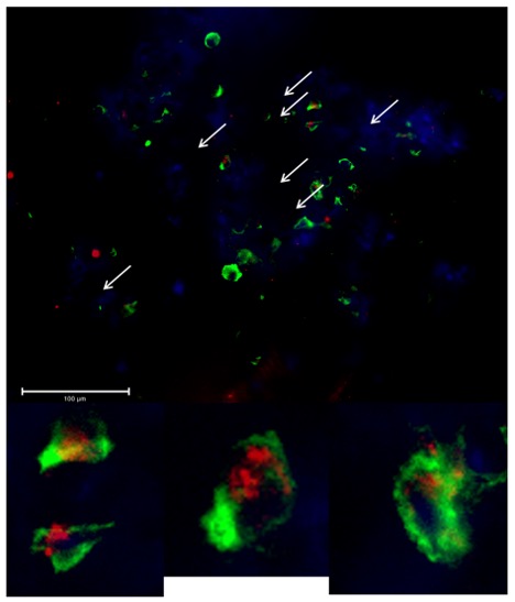
*Results: Hydrodynamic size of anti-IL6R-Alexa594 NP was 107 ± 14 nm (polydispersity index: 0.25), transmission electron microscopy size was 98 ± 8.1 nm, and zeta-potential was -3.1 ± 1.2 mV. We confirmed loading efficiency of anti-IL6R-mAb in NP as 23.1 ± 3.8% using bicinchoninic assay. Anti-IL6R-Alexa594 NP showed 50% of drug elution by 4.6 days and 75% of drug elution by 13 days . Anti-IL6R-Alexa594 NP were found in the lung parenchyma and in the hilar lymph nodes. Immunofluorescence staining assay demonstrated that CD163+, CD11b+, HLADR+, and CD11c+ cells from hilar lymph node internalized NPs. The same held true for the lung parenchyma tissue with the addition of CD3+ cells also having nanoparticle uptake. Approximately 10% of the lymph node macrophages (HLADR+, CD11b+, CD163+) and 50% of the pulmonary macrophages had NP uptake. Figure 1: Immunofluorescence imaging of lung parenchyma. Blue = DAPI, Green = HLADR+, Red = NP.
*Conclusions: In this study, we synthesized bright nanovesicles to track the fate of NPs in the lung. These biocompatible NP produced sustained release of their loaded anti-IL6R over the span of two weeks in physiological condition, offering a way to control immune reactions locally. Despite the short perfusion time, a significant number of lung alveolar macrophages were able to phagocytose NPs. This is a promising new avenue for drug delivery in hopes of reducing incidence and severity of lung PGD.
To cite this abstract in AMA style:
Patel PM, Jung S, Miller CL, O JM, Costa T, Dehnadi A, Hanekamp I, Li XF, Jiang L, Ichimura H, Azimzadeh A, Madsen JC, Abdi R. Nanoparticle Mediated Drug Delivery for Lung Transplantation [abstract]. Am J Transplant. 2021; 21 (suppl 3). https://atcmeetingabstracts.com/abstract/nanoparticle-mediated-drug-delivery-for-lung-transplantation/. Accessed January 2, 2026.« Back to 2021 American Transplant Congress

