Application of Diffusional Kurtosis Imaging for Non-Invasive Assessment of Chronic Allograft Injury
1Department of Urology, Capital Medical University Beijing Chaoyang Hospital, Urology Institute of Capital Medical University, Beijing, China, 2Urology Department, Beijing YouAn Hospital, Capital Medical University, Beijing, China
Meeting: 2020 American Transplant Congress
Abstract number: B-092
Keywords: Graft function, Kidney transplantation, Magnetic resonance imaging
Session Information
Session Name: Poster Session B: Kidney Complications: Non-Immune Mediated Late Graft Failure
Session Type: Poster Session
Date: Saturday, May 30, 2020
Session Time: 3:15pm-4:00pm
 Presentation Time: 3:30pm-4:00pm
Location: Virtual
*Purpose: To evaluate the feasibility and clinical application of diffusional kurtosis imaging (DKI) technique in non-invasive assessment for chronic allograft injury (CAI).
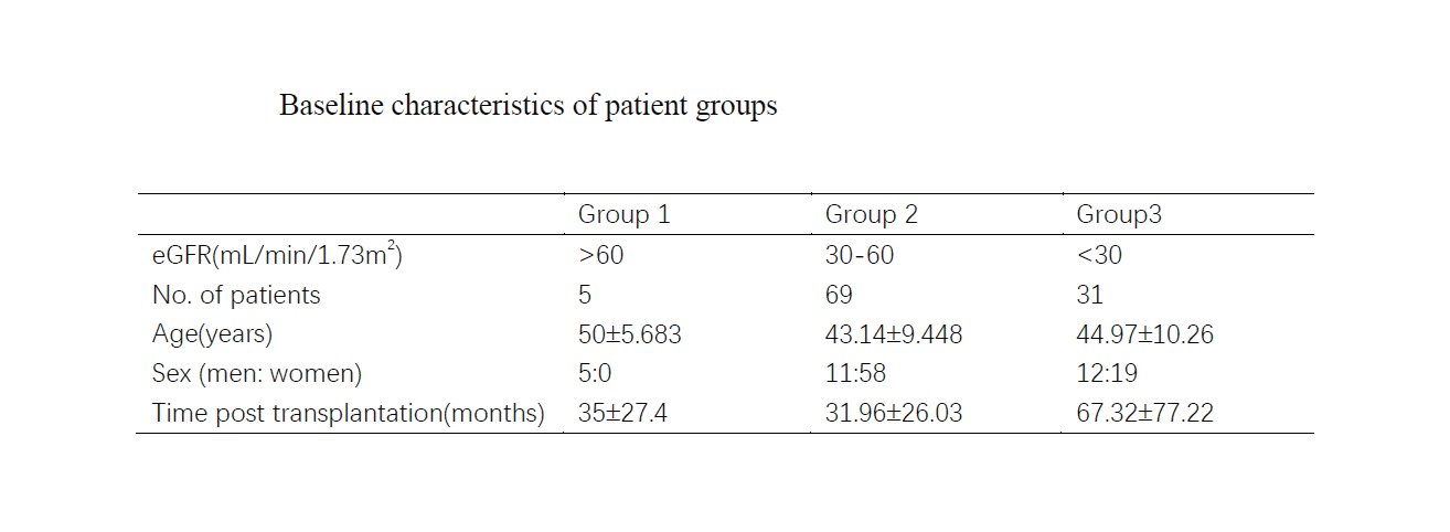
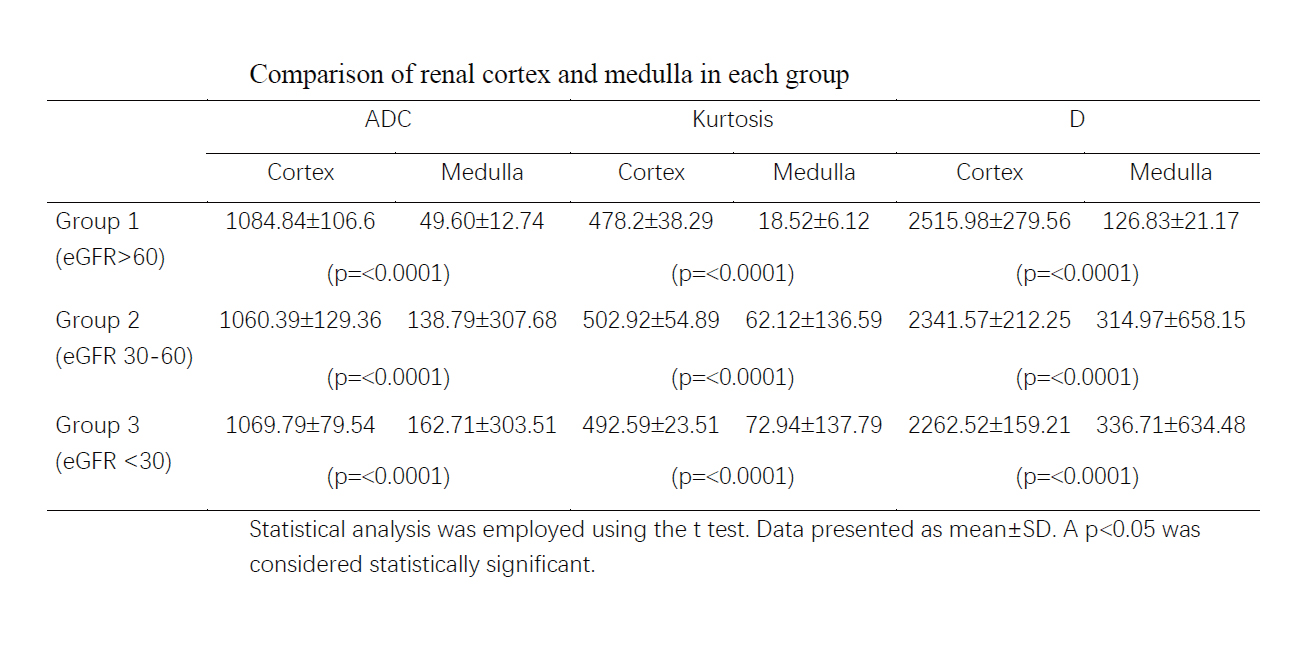
*Methods: The local ethics committee approved this study; written informed consent was obtained. Between January 2018 and October 2019, a total of 105 renal allograft recipients were recruited and divided into allograft function normal(eGFR≥60) and abnormal(eGFR<60) groups for analysis and screening of relevant DKI parameters. Then they were grouped on eGFR (mL/min/1.73m2) levels to 3 groups: group 1, eGFR≥60(n=5); group 2, eGFR 30-60 (n=69); group 3, eGFR<30 (n=31). DKI was performed on a clinical 3 T magnetic resonance imaging (MRI) system, and region-of-interest measurements were conducted to determine mean kurtosis (K), mean diffusivity (D), and apparent diffusion coefficient (ADC) of the kidney cortex and medulla. Renal biopsy specimens were performed in allograft functionabnormal group randomly.
*Results: Mean D values of the renal cortex were significantly lower in the eGFR abnormal group. And Mean D values of the renal cortex decreased gradually with the increase of creatinine, whereas there is a significant difference in the value of D values of cortex between different eGFR groups. (p<0.05). And biopsy result confirmed interstitial fibrosis and tubular atrophy (IFTA).
*Conclusions: As allograft injury progresses, the perfusion of renal might be reduced, and the molecular of the renal cortex might be influenced. The DKI quantitative parameter D of the renal cortex can non-invasively assess CAI to some extent. DKI technique is expected to be an effective, natural, and non-invasive method to detect CAI.
To cite this abstract in AMA style:
Xu Y, Zheng X, Liu H, Hu X. Application of Diffusional Kurtosis Imaging for Non-Invasive Assessment of Chronic Allograft Injury [abstract]. Am J Transplant. 2020; 20 (suppl 3). https://atcmeetingabstracts.com/abstract/application-of-diffusional-kurtosis-imaging-for-non-invasive-assessment-of-chronic-allograft-injury/. Accessed February 22, 2026.« Back to 2020 American Transplant Congress


