Alterations in Cerebral Blood Flow before and after Kidney Transplantation
A. Gupta1, I. Choi2, R. Lepping3, P. Adany4, A. Jurgensen1, J. Klein1, D. Cibrik1, J. Burns5, W. Brooks2, P. Lee2
1Division of Nephrology, The Kidney Institute, University of Kansas Medical Center, Kansas City, KS, 2Hoglund Brain Imaging Center, Department of Molecular & Integrative Physiology, University of Kansas Medical Center, Kansas City, KS, 3Hoglund Brain Imaging Center, Alzheimer’s Disease Center,, University of Kansas Medical Center, Kansas City, KS, 4Hoglund Brain Imaging Center, University of Kansas Medical Center, Kansas City, KS, 5Alzheimer’s Disease Center, Department of Neurology, University of Kansas Medical Center, Kansas City, KS
Meeting: 2019 American Transplant Congress
Abstract number: C197
Keywords: Calcineurin, Kidney/liver transplantation, Risk factors
Session Information
Session Name: Poster Session C: Kidney: Cardiovascular and Metabolic
Session Type: Poster Session
Date: Monday, June 3, 2019
Session Time: 6:00pm-7:00pm
 Presentation Time: 6:00pm-7:00pm
Location: Hall C & D
*Purpose: End stage renal disease (ESRD) is associated with alterations in cerebral blood flow (CBF) likely secondary to disrupted cerebral autoregulation. In this study we examined whether CBF normalizes after a successful kidney transplant (KT).
*Methods: Pre-KT ESRD patients underwent brain magnetic resonance imaging with arterial spin labeling (ASL) to detect regional CBF in 17 regions of the brain before and 12 weeks after KT. CBF data were processed using a customized data-processing pipeline and corrected for hematocrit levels. We compared CBF between pre-KT, post-KT, and healthy controls using t-tests. All patients were on calcineurin inhibitors post-KT.
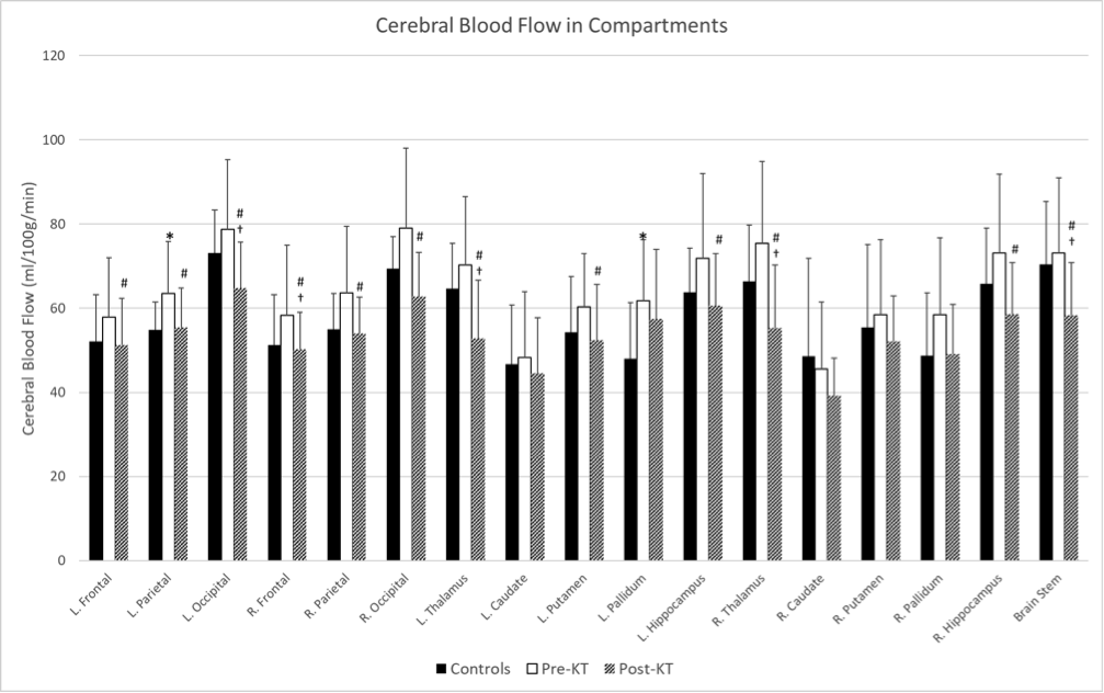
*Results: We analyzed CBF in 11 healthy controls (age 54 ± 7, 5/11 men, 10/11 Caucasians) and 17 patients with ESRD (age 50 ± 9, 12/17 men, 16/17 Caucasians). Figure 1 shows the CBF in controls, and pre- and post-KT ESRD in the various brain regions (*: p < 0.05 for pre-KT vs. controls; †: p < 0.05 for post-KT vs. controls; #: P<0.01 for pre-KT vs. post-KT). Pre-KT CBF showed a trend toward higher values than controls. Post-KT CBF was significantly reduced compared to pre-KT CBF and even lower than controls in five brain regions including the frontal and occipital lobes, thalamus, and brain stem.
*Conclusions: KT normalized the increase in CBF in kidney disease and further reduced CBF in certain brain regions below controls. Further studies are needed to understand whether this post-KT decrease in CBF is secondary to improvement in cerebral autoregulation or due to cerebral vasoconstriction due to calcineurin inhibitors.
To cite this abstract in AMA style:
Gupta A, Choi I, Lepping R, Adany P, Jurgensen A, Klein J, Cibrik D, Burns J, Brooks W, Lee P. Alterations in Cerebral Blood Flow before and after Kidney Transplantation [abstract]. Am J Transplant. 2019; 19 (suppl 3). https://atcmeetingabstracts.com/abstract/alterations-in-cerebral-blood-flow-before-and-after-kidney-transplantation/. Accessed February 1, 2026.« Back to 2019 American Transplant Congress

