Alterations in Brain Metabolites before and after Kidney Transplant
A. Gupta1, P. Lee2, R. Lepping3, A. Jurgensen1, J. Klein1, P. Adany4, D. Cibrik1, J. Burns5, W. Brooks3, I. Choi6
1Division of Nephrology, The Kidney Institute, University of Kansas Medical Center, Kansas City, KS, 2Hoglund Brain Imaging Center, Department of Molecular & Integrative Physiology, University of Kansas Medical Center, Kansas City, KS, 3Hoglund Brain Imaging Center, Alzheimer’s Disease Center,, University of Kansas Medical Center, Kansas City, KS, 4Hoglund Brain Imaging Center, University of Kansas Medical Center, Kansas City, KS, 5Alzheimer’s Disease Center, Department of Neurology, University of Kansas Medical Center, Kansas City, KS, 6Hoglund Brain Imaging Center, Department of Neurology, University of Kansas Medical Center, Kansas City, KS
Meeting: 2019 American Transplant Congress
Abstract number: C188
Keywords: Image analysis, Kidney transplantation, Morbidity, Risk factors
Session Information
Session Name: Poster Session C: Kidney: Cardiovascular and Metabolic
Session Type: Poster Session
Date: Monday, June 3, 2019
Session Time: 6:00pm-7:00pm
 Presentation Time: 6:00pm-7:00pm
Location: Hall C & D
*Purpose: End stage renal disease (ESRD) is associated with accumulation of uremic metabolites and poor cognitive performance. Alterations in brain metabolites are associated with cognitive impairment in several disease states. We examined whether the brain metabolites were altered in ESRD and whether they normalize post- kidney transplant (KT).
*Methods: We analyzed brain metabolites pre- and 12 weeks post- KT. We used magnetic resonance spectroscopic imaging (MRS) at 3 Tesla (Skyra, Siemens) to measure N-acetylaspartate (NAA), choline (Cho), creatine (Cr), myo-inositol (mI), and glutamate and glutamine (Glx) in a single slice of brain superior to the lateral ventricles. Brain metabolite signals were quantified using LCModel and reported as ratios to creatine for normalization. Comparisons of metabolite concentrations between pre- and post-KT, and ESRD and controls concentrations were made using a paired t-test.
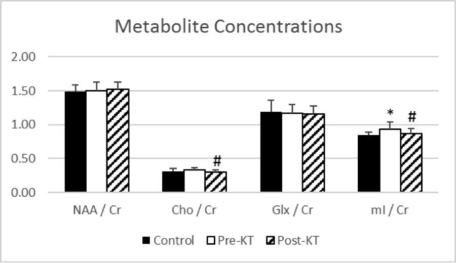
*Results: MRS data from 11 healthy controls (age 54 ± 7, 5/11 men, 10/11 Caucasians) and 18 patients with ESRD (age 51 ± 11, 13 men, 17 Caucasians) were compared. Figure 1 shows the metabolite concentrations in controls, pre-KT and post-KT patients (*: P< 0.05 for pre-KT vs. controls; #: P< 0.05 pre-KT vs. post-KT). The mI/Cr ratio was elevated pre-KT when compared to healthy controls (P= 0.03) and decreased by 7% post-KT (P =0.008). In addition, Cho/Cr ratio was numerically higher in pre-KT ESRD and decreased by 10% post-KT (P <0.001). There was no difference in metabolites between controls and post-KT patients. There were no tissue composition (gray and white matter) differences between the analyzed brain regions.
*Conclusions: Kidney disease is associated with specific metabolic disturbances in the brain that normalize after KT. These improvements may account for improvement in cerebrovascular health and cognition function post-transplant.
To cite this abstract in AMA style:
Gupta A, Lee P, Lepping R, Jurgensen A, Klein J, Adany P, Cibrik D, Burns J, Brooks W, Choi I. Alterations in Brain Metabolites before and after Kidney Transplant [abstract]. Am J Transplant. 2019; 19 (suppl 3). https://atcmeetingabstracts.com/abstract/alterations-in-brain-metabolites-before-and-after-kidney-transplant/. Accessed January 6, 2026.« Back to 2019 American Transplant Congress

