Kidney Transplantation is Associated with Decreasing Native T1 Time: A Cardiac Magnetic Resonance Study
M. Contti,1 L. de Andrade,1 H. Nga,1 M. Valiatti,1 H. Takase,1 A. Bravin,1 A. Mauricio,2 M. Barbosa.3
1Internal Medicine, Botucatu Medical School, Botucatu, Sao Paulo, Brazil
2Radiology, Heart Institute of the Clinics Hospital at the Faculty of Medicine, University of São Paulo, São Paulo, Brazil
3Radiology, Centro Avançado de Radiologia e Diagnóstico por Imagem, Presidente Prudente, Sao Paulo, Brazil.
Meeting: 2018 American Transplant Congress
Abstract number: A209
Keywords: Magnetic resonance imaging, Non-invasive diagnosis, Radiologic assessment, Risk factors
Session Information
Session Name: Poster Session A: Kidney: Cardiovascular and Metabolic
Session Type: Poster Session
Date: Saturday, June 2, 2018
Session Time: 5:30pm-7:30pm
 Presentation Time: 5:30pm-7:30pm
Location: Hall 4EF
Patients with stage 5 chronic kidney disease (CKD) have high rate of cardiovascular death. Kidney transplantantion, on the other hand, is associated with lower cardiovascular morbidity and mortality. Cardiac magnetic resonance (CMR) is the “gold-standard” for measureament ventricular dimensions. Recently, native T1 mapping has been proposed as a contrast-free method of fibrosis assessment. There is currently no study evaluating native T1 following kidney transplantation.
PURPOSE: Avaliate the change of native T1 times by CMR 6 months after kidney transplantation.
METHODS: Forty-four kidney transplant patients underwent two CMR scans: the first one, within the first 10 days of procedure; and the second one, 6 months thereafter. In addition to the native T1, interventricular septum, left ventricular posterior wall thickness, left ventricular mass, left ventricular mass index, were evaluated. Likewise, left and right ventricular parameters has been measured: ejection fraction, end-systolic volume, end-diastolic volume, end-systolic volume index, end-diastolic volume index, ejection volume and ejection volume index.
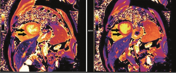
RESULTS: A statistically significant reduction was observed in native T1 times, from 1331 (± 52) ms to 1298 (± 42) ms 6 months after transplantation, p = 0.001. Figure 1 shows the scan of a patient with a significant reduction in T1 time. Left and right ventricular parameters presented no difference between the following moments of the exams. 
CONCLUSIONS: This study is the first to evaluate native T1 mapping in kidney transplant patients. It showed that kidney transplantation, even in the short period of 6 months, is associated with a significant reduction of native T1 in CMR, probably reducing myocardial fibrosis.
CITATION INFORMATION: Contti M., de Andrade L., Nga H., Valiatti M., Takase H., Bravin A., Mauricio A., Barbosa M. Kidney Transplantation is Associated with Decreasing Native T1 Time: A Cardiac Magnetic Resonance Study Am J Transplant. 2017;17 (suppl 3).
To cite this abstract in AMA style:
Contti M, Andrade Lde, Nga H, Valiatti M, Takase H, Bravin A, Mauricio A, Barbosa M. Kidney Transplantation is Associated with Decreasing Native T1 Time: A Cardiac Magnetic Resonance Study [abstract]. https://atcmeetingabstracts.com/abstract/kidney-transplantation-is-associated-with-decreasing-native-t1-time-a-cardiac-magnetic-resonance-study/. Accessed January 27, 2026.« Back to 2018 American Transplant Congress
