In Vivo Imaging and Function of Renal Tertiary Lymphoid Organs
Starzl Transplantation Institute, Pittsburgh.
Meeting: 2018 American Transplant Congress
Abstract number: 10
Keywords: Antigen presentation, Kidney, Mice, T cell activation
Session Information
Session Name: Concurrent Session: Antigen Presentation / Allorecognition / Dendritic Cells
Session Type: Concurrent Session
Date: Sunday, June 3, 2018
Session Time: 2:30pm-4:00pm
 Presentation Time: 3:30pm-3:42pm
Location: Room 4C-3
Tertiary lymphoid organs (TLOs) are ectopic lymphoid structures that arise in non-lymphoid tissues in the setting of chronic inflammation such as organ transplantation. The function of TLOs in transplantation is controversial, with studies demonstrating a role in rejection as well as in immune regulation. Therefore, fundamental understanding of TLO function is necessary. Here, we employed intravital time-lapse 2-photon microscopy to directly visualize cellular interactions within TLOs that form under the kidney capsule in RIP-LTα mice.
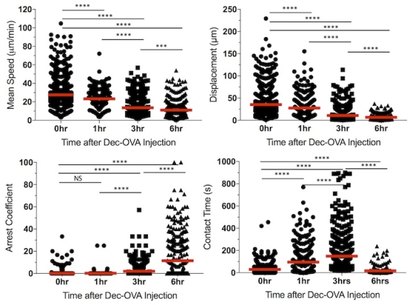
B6-RIPLTα CD11c-YFP BM chimeric mice were adoptively transferred with 10e6 naïve dsRed OT-I T cells and imaged 24 hours later. Evans Blue and MadCAM-1-PE mAb were used i.v. to visualize blood flow and high endothelial venules, respectively. TLOs were identified by the presence of discrete T and dendritic cell (DC) accumulations, lack of typical kidney architecture, and positive MadCAM-1 staining. To visualize T cell kinetics after antigen exposure, imaging was performed at hrs 0 (no Ag exposure), 1, 3, and 6 after mice were injected with DEC205-OVA fusion antibody and CD40 mAb (FGK4.5). Three-dimensional image analysis was performed and OT-I T cell mean speed, displacement, arrest coefficient, and contact time with DCs were calculated. Mean speed and displacement significantly decreased over time upon antigen exposure while arrest coefficient and mean contact time significantly increased. These data are consistent with productive T cell-DC interactions and mirror previously reported data in secondary lymphoid organs.
Using dynamic intravital imaging, we are presenting direct evidence that naïve T cells traffic to TLOs but not surrounding non-lymphoid tissue, and have productive interactions with DCs when antigen is present. We have established a model that will be utilized to study the interactions of immune cells in TLOs in real time. This will allow us to investigate T cell, B cell, and regulatory T cell function in TLOs in the setting of mouse kidney transplantation. The findings will elucidate the role of TLO in transplantation and will be relevant to other settings of chronic inflammation such as autoimmune disease, cancer, and chronic infection.
CITATION INFORMATION: Abou-Daya K., Zhao D., Williams A., Oberbarnscheidt M. In Vivo Imaging and Function of Renal Tertiary Lymphoid Organs Am J Transplant. 2017;17 (suppl 3).
To cite this abstract in AMA style:
Abou-Daya K, Zhao D, Williams A, Oberbarnscheidt M. In Vivo Imaging and Function of Renal Tertiary Lymphoid Organs [abstract]. https://atcmeetingabstracts.com/abstract/in-vivo-imaging-and-function-of-renal-tertiary-lymphoid-organs/. Accessed February 13, 2026.« Back to 2018 American Transplant Congress

