Imaging Cell Biology In Vivo with Intravital-Microscopy.
1Renal Division, Transplantation Research Center, Brigham and Women's Hospital, Boston, MA
2Transplant Surgery, Tokyo Medical Univ., Hachioji, Tokyo, Japan
3Intensive Care Medicine, Tokyo Medical Univ., Hachioji, Tokyo, Japan.
Meeting: 2016 American Transplant Congress
Abstract number: B4
Keywords: Antigen presentation, Co-stimulation, Image analysis, Mice
Session Information
Session Name: Poster Session B: Allograft Rejection, Tolerance, and Xenotransplantation
Session Type: Poster Session
Date: Sunday, June 12, 2016
Session Time: 6:00pm-7:00pm
 Presentation Time: 6:00pm-7:00pm
Location: Halls C&D
Background: Despite current immunosuppressive therapy, many recipients endure multiple episodes of acute rejection. The interplay between dendritic cells (DCs) and T cells plays a central role in the process of allograft rejection, however, little is known about the temporal and spatial interplay of these events. Recently, we showed that graft DC-SIGN+ cells correlated with poor allograft survival and with allograft inflammation 1. To develop novel strategies designed to achieve durable tolerance and prevent chronic rejection, our research is aimed at understanding of mechanisms of " the interplay – when, where, and how ..".
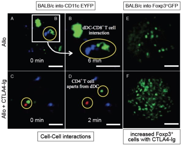
Methods: We used a murine cardiac transplant model: isograft (B6 heart into CD11c EYFP B6 recipient) and allograft (BALB/c heart) -/+ CTLA4-Ig (500 [mu]g Day 0, 250 [mu]g Day 2,4). To investigate beneficial effects of CTLA4-Ig in allograft, we also used BALB/c heart into Foxp3+ GFP B6 recipient -/+ CTLA4-Ig model. Recipients were received 5millions of CD4 (labeled as red) or CD8 (blue) i.v. injections right after surgery to see cellular interactions with using a novel in vivo imaging tool 2. We examined: 1. DCs morphology & numbers, 2. DCs-T cell interplay, 3. Regulation of Tregs.
Results: Administration of CTLA4-Ig in allograft resulted in better DC regulation in morphology , less antigation presentation, shorter period of interplay between DC and T cell, and increase of Tregs compared to control.
Conclusions: We investigated the role of recipient DC-T cell interaction in graft rejection versus tolerance utilizing state-of-the-art imaging technologies. Dynamic images from current advanced in vivo imaging tools represent as a powerful tool to provide quantitative and dynamic insights into transplant immunology and will serve as a foundation for protective strategies focused on promoting tolerance.
References: 1. Batal I, et al. J Am Soc Nephrol. 2015, 2. Jung K, et al. Circ Res. 2013

CITATION INFORMATION: Ueno T, Yeung M, Igarashi N, Yokoyama T, Kihara Y, Konno O, Nakamura Y, Iwamoto H, Ikeda T, McGrath M, Sayegh M, Chandraker A. Imaging Cell Biology In Vivo with Intravital-Microscopy. Am J Transplant. 2016;16 (suppl 3).
To cite this abstract in AMA style:
Ueno T, Yeung M, Igarashi N, Yokoyama T, Kihara Y, Konno O, Nakamura Y, Iwamoto H, Ikeda T, McGrath M, Sayegh M, Chandraker A. Imaging Cell Biology In Vivo with Intravital-Microscopy. [abstract]. Am J Transplant. 2016; 16 (suppl 3). https://atcmeetingabstracts.com/abstract/imaging-cell-biology-in-vivo-with-intravital-microscopy/. Accessed January 14, 2026.« Back to 2016 American Transplant Congress
