Computed Tomography Measures of Nutrition Deficits in Patients with Intestine Failure.
Surgery, Transplant Division, Indiana University School of Medicine, Indianapolis, IN.
Meeting: 2016 American Transplant Congress
Abstract number: A298
Keywords: Intestinal transplantation
Session Information
Session Name: Poster Session A: Small Bowel: All Topics
Session Type: Poster Session
Date: Saturday, June 11, 2016
Session Time: 5:30pm-7:30pm
 Presentation Time: 5:30pm-7:30pm
Location: Halls C&D
Introduction: Patients with intestine failure (IF) and dependence on parenteral nutrition (PN) develop severe nutrition deficits that impact on perioperative morbidity and mortality. Previous research demonstrates that laboratory measures of nutrition status fail to assess clinical deficits in muscle mass and fat tissue. This study employs computed tomography imaging to assess (1) muscle mass and (2) subcutaneous and (3) visceral fat stores in patients with long standing IF.
Methods: A 1:1 case-control study design is used to compare IF patients with healthy controls. atients were selected from a database of IF patients, known to have long-standing PN dependence and being evaluated for isolated intestine transplant. Controls were selected from the trauma database, as patients with an injury severity score less than 10, who received an abdominal CT scan. Cases and controls were matched for age +/- 5 years, gender, and BMI +/- 2.
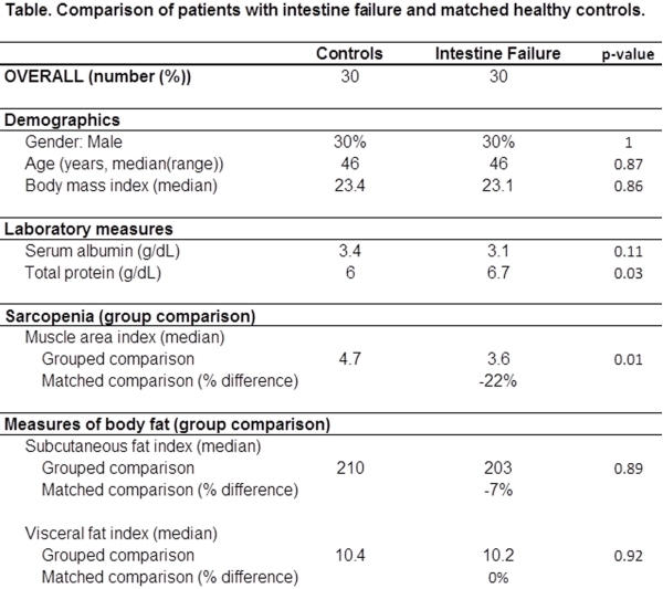
Results: There were 30 subjects and 30 controls. Compared to healthy controls, the IF patients had a higher serum protein level (6.7 vs 6.0, p=0.03), but a lower serum albumin level (3.1 vs 3.4, p=0.11). The IF patients had a -22% muscle mass deficit and a -7% subcutaneous fat deficit, but there was no difference in visceral fat stores. Subgroup analysis by gender demonstrated a greater loss of muscle mass in IF males compared to IF females, (-36% vs -16%, p=0.26); a similar deficit in subcutaneous fat (-9% IF male vs -6% IF female, p=0.86); and a greater loss of visceral fat for males (-14% vs +6%, p=0.45). Three age groups were analyzed: < 40, 40-60, and >60 years. These three group had incrementally greater deficit of muscle mass (-42%, -25%, +12%) with lower age (p=0.03). The age groups did not differ significantly for fat deficits.
Conclusions: These results support previous research demonstrating substantial nutritional deficits in IF patients. Standard CT scans can be employed to assess the distribution and quantity of nutritional deficits. IF patients in this study have significant deficits of muscle and subcutaneous fat stores, but a similar amount of visceral fat. These findings vary by age and gender.

CITATION INFORMATION: Davis J, Bush W, Kubal C, Mangus R. Computed Tomography Measures of Nutrition Deficits in Patients with Intestine Failure. Am J Transplant. 2016;16 (suppl 3).
To cite this abstract in AMA style:
Davis J, Bush W, Kubal C, Mangus R. Computed Tomography Measures of Nutrition Deficits in Patients with Intestine Failure. [abstract]. Am J Transplant. 2016; 16 (suppl 3). https://atcmeetingabstracts.com/abstract/computed-tomography-measures-of-nutrition-deficits-in-patients-with-intestine-failure/. Accessed February 27, 2026.« Back to 2016 American Transplant Congress
