Successful Kidney Transplantation in a Small Child with an Atretic Inferior Vena Cava
1Michael E. DeBakey Department of Surgery, Baylor College of Medicine, Houston, TX, 2Department of Pediatrics, Baylor College of Medicine, Houston, TX
Meeting: 2019 American Transplant Congress
Abstract number: C245
Keywords: Kidney transplantation, Outcome, Pediatric, Surgical complications
Session Information
Session Name: Poster Session C: Kidney: Pediatrics
Session Type: Poster Session
Date: Monday, June 3, 2019
Session Time: 6:00pm-7:00pm
 Presentation Time: 6:00pm-7:00pm
Location: Hall C & D
*Purpose: Kidney transplantation is the treatment of choice in pediatric patients with end stage renal disease. This population presents technical challenges particularly in those less than 20 kgs due to anomalous anatomy, vascular access issues prior to transplantation, and a generally small size for age. Herein, we report a case of a pediatric kidney transplantation where successful allograft outflow was achieved using the superior mesenteric vein (SMV) when he was found to have an atretic inferior vena cava (IVC) intraoperatively.
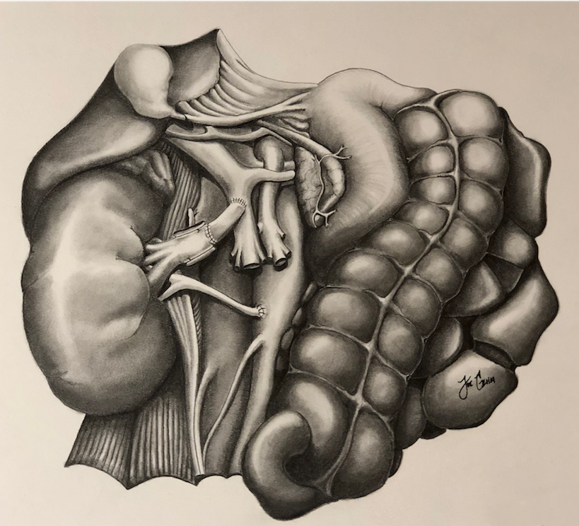
*Methods: Standard allograft outflow is usually achieved utilizing the iliac veins or IVC. When use of the iliocaval system is not feasible, alternative anastomosis must be considered. In this case, we created a donor iliac graft for added length to anastomose the renal vein with the SMV. The SMV and portal vein were dissected from below the pancreas and exposed. A side-biting vascular clamp was used and the donor Iliac graft was anastomosed to the SMV proximal to the coalescence of the portal vein with the splenic. The iliac vein graft provided sufficient length and was found to be of adequate size in comparison to the donor renal vein [figure1]. Arterial anastomosis was performed in an end-to-side fashion from the renal artery directly to the infrarenal aorta.
*Results: In the setting of IVC occlusion with poor drainage, we utilized a patent vessel with larger caliber for outflow to reduce the risk of high venous pressures, allograft failure, venous rotation, and thrombosis. Reperfusion of the allograft was uneventful the second time, with the allograft appearing perfused with excellent color, turgor and pulse along the renal artery. An intraoperative ultrasound confirmed patent renal vessels with adequate flow throughout the vessels, including the renal vein conduit.
*Conclusions: We conclude that the SMV may serve as an alternative outflow tract in the small pediatric patient and provides the vessel caliber needed to reduce the risks of complications. This approach has shown to be a safe and effective way to provide sufficient outflow in the setting of IVC occlusion in the pediatric recipient
To cite this abstract in AMA style:
Geha JA, Geha JD, Goss M, Kueht ML, Cotton RT, Goss JA, Rana A, O'Mahony CA, Srivaths P, Brewer E, Galvan NN. Successful Kidney Transplantation in a Small Child with an Atretic Inferior Vena Cava [abstract]. Am J Transplant. 2019; 19 (suppl 3). https://atcmeetingabstracts.com/abstract/successful-kidney-transplantation-in-a-small-child-with-an-atretic-inferior-vena-cava/. Accessed February 15, 2026.« Back to 2019 American Transplant Congress

