Real-Time Three-Dimensional Fourier-Domain Optical Coherence Tomography for Intravital Imaging of Thrombosis after Vascularized Composite Allotransplantation Procedures
Department of Plastic and Reconstructive Surgery, Johns Hopkins University School of Medicine, Baltimore, MD
Department of Surgery, Jinling Hospital, Nanjing University School of Medicine, Nanjing, Jiangsu, China
Department of Electrical and Computer Engineering, Johns Hopkins University, Baltimore, MD
Meeting: 2013 American Transplant Congress
Abstract number: A849
Purpose: We sought to apply real-time three-dimensional Fourier-domain optical coherence tomography (3D FD-OCT) for in vivo monitoring of vascular thrombosis and thrombolysis.
Methods: 10% Ferric chloride (FeCl3) solution was used to induce thrombus formation within the femoral artery (FA). 3D FD-OCT running at 70,000 A-scans per second with lateral resolution of 12 ¯o;m and axial resolution of 3.6 ¯o;m was used for intravital imaging at serial time points and after thrombus formation. Low-molecular-weight heparin (200 U/kg) was injected i.v. after early stage of thrombus formation and detection by 3D FD-OCT. Experimental vessels were harvested for hematoxylin and eosin (H&E) staining.
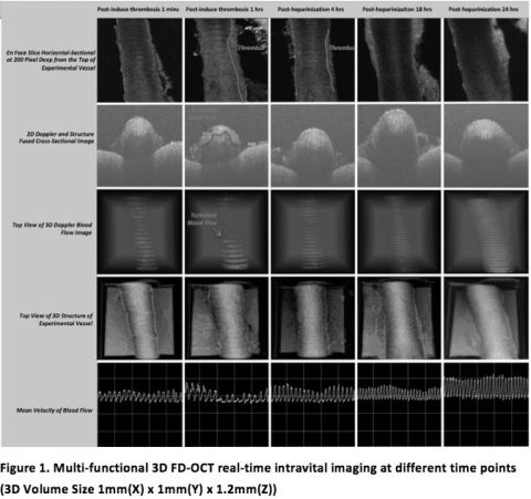
Results: Femoral arterial thrombus commenced to form at 4 min after initiation of FeCl3-induced injury of the FA and complete vessel occlusion occurred at around 12 min. We were able demonstrate that 3D FD-OCT allowed to monitor the whole pathophysiological cascade of thrombogenesis, detect partial thrombi that were not detectable under traditional microscopy, and assess the effect of anticoagulation treatment until complete resolution of the thrombus was achieved by 2D tomographic slides, 3D anatomic and Doppler image, and mean velocities of blood flow.

Thrombus formation was confirmed by H&E histology and correlated with 3D FD-OCT imaging.
Conclusion: 3D FD-OCT enables assessment of arterial thrombosis and thrombolysis in real-time, non-invasively and with three-dimensional high resolution. 3D FD-OCT can be used for early intraoperative detection of thrombus formation and to monitor the effect of anticoagulation. This could ultimately aid to intraoperative management and help to reduce complications and patient morbidity after VCA.
To cite this abstract in AMA style:
Mao Q, Huang Y, Pang J, Zhu S, Tong D, Ibrahim Z, Christensen J, Li Y, Li J, Lee W, Kang J, Brandacher G. Real-Time Three-Dimensional Fourier-Domain Optical Coherence Tomography for Intravital Imaging of Thrombosis after Vascularized Composite Allotransplantation Procedures [abstract]. Am J Transplant. 2013; 13 (suppl 5). https://atcmeetingabstracts.com/abstract/real-time-three-dimensional-fourier-domain-optical-coherence-tomography-for-intravital-imaging-of-thrombosis-after-vascularized-composite-allotransplantation-procedures/. Accessed February 26, 2026.« Back to 2013 American Transplant Congress
