More Than Meets the Eye: The Use of Serial Intravascular Ultrasound for Assessment of Cardiac Allograft Vasculopathy after Heart Transplantation
1Stanford University, Palo Alto, CA, 2University of Toronto, Toronto, ON, Canada
Meeting: 2019 American Transplant Congress
Abstract number: 94
Keywords: Angiography, Arteriosclerosis, Intimal, Prediction models
Session Information
Session Name: Concurrent Session: Medley of Heart Transplantation Topics
Session Type: Concurrent Session
Date: Sunday, June 2, 2019
Session Time: 2:30pm-4:00pm
 Presentation Time: 3:30pm-3:42pm
Location: Room 206
*Purpose: Cardiac allograft vasculopathy (CAV) is the “Achilles heel” of long-term survival after heart transplantation. While conventional coronary angiography is recognised as the standard for the diagnosis of CAV, it is insensitive for early and diffuse disease compared to intravascular ultrasound (IVUS). An increase in maximal intimal thickness (MIT) ≥ 0.5 mm in the first year is associated with an increased risk of subsequent death but there is limited data for determining clinically relevant changes in MIT after the first year. We evaluated serial IVUS in patients with and without CAV at a large heart transplant center.
*Methods: This was a retrospective single center study of all adult heart transplant patients (2008-2017) with serial IVUS at baseline and annually for 5 years was completed. Hidden Markov Models (HMMs) were developed. We applied a two-state HMM (angiographic CAV and CAV-free) to analyze observed pairs of MITs and angiography-based assessments and to assess progression of disease over time. We estimated the distribution of MIT conditional on disease status as well as the transition of probabilities for 5 years. Based on the inferred disease status, we assessed the incremental MIT change associated with CAV using linear mixed models.
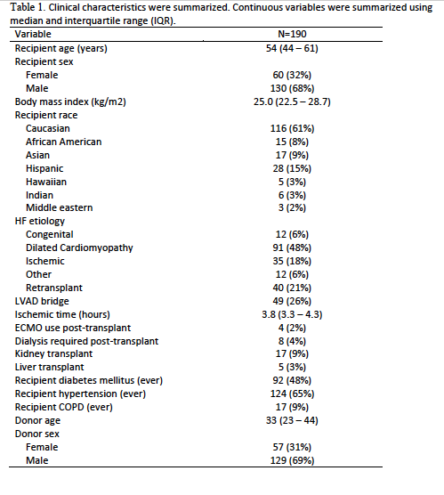
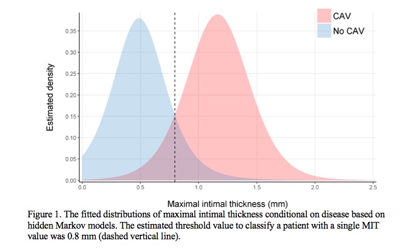
*Results: Among 284 recipients screened, 190 (67%) had at least 2 IVUS measurements within 5 years. Baseline characteristics are summarized in Table 1. The estimated mean (SD) MIT was 0.5 (±0.3) mm in those without CAV. The threshold value to classify a patient with CAV based on a single MIT was 0.8 mm (the dashed vertical line) on Figure 1. Compared to those remaining CAV-free at year 1, an increase in MIT of 0.64 mm (p<0.001) was the threshold for development of CAV at year 2. Subsequently, an MIT increase of 0.62 mm + 0.02 * (years between 2 IVUS studies) determined the relevant change needed for the development of angiographic CAV at any given time.
*Conclusions: Among heart transplant patients undergoing IVUS for assessment of CAV, an absolute MIT of 0.8 mm differentiated those with and without CAV at any time. An MIT increase of 0.64 mm at year 1, 0.66 mm between years 1 and 3 and 0.70 mm between years 1 to 5 was associated with the development of CAV.
To cite this abstract in AMA style:
Moayedi Y, Fan C, Tremblay-Gravel M, Miller RJ, Kawana M, Parizo J, Oro G, Wainwright R, Luikart HI, D'Emilio N, Gordon J, Kameda R, Ikutomi M, Manlhiot C, Fearon WF, Ross HJ, Khush KK, Teuteberg JJ. More Than Meets the Eye: The Use of Serial Intravascular Ultrasound for Assessment of Cardiac Allograft Vasculopathy after Heart Transplantation [abstract]. Am J Transplant. 2019; 19 (suppl 3). https://atcmeetingabstracts.com/abstract/more-than-meets-the-eye-the-use-of-serial-intravascular-ultrasound-for-assessment-of-cardiac-allograft-vasculopathy-after-heart-transplantation/. Accessed February 10, 2026.« Back to 2019 American Transplant Congress


