Impact of Abdominal Aortic Calcification in Living Kidney Donors
1Department of Urology, Yonsei University College of Medicine, Seoul, Korea
2Department of Urology, CHA University, Seongnam-si, Korea
3Department of Transplantation Surgery, Yonsei University College of Medicine, Seoul, Korea.
Meeting: 2015 American Transplant Congress
Abstract number: 299
Keywords: Arteriosclerosis, Donation, Kidney transplantation, Renal function
Session Information
Session Name: Concurrent Session: Kidney: Living Donor Issues II
Session Type: Concurrent Session
Date: Monday, May 4, 2015
Session Time: 4:00pm-5:30pm
 Presentation Time: 4:24pm-4:36pm
Location: Terrace IV
Introduction: To assess the association between abdominal aortic calcification (AAC) and renal function of living kidney donor and to evaluate the value of an AAC score as a surrogate marker for the presence of nephrosclerosis.
Methods: Between January 2010 and March 2013, 287 donors who underwent live donor nephrectomy were enrolled. CT angiographies were analyzed and calcification score (CS) was quantified by calculating Agatston score in abdominal aorta. The donors were stratified into a non-AAC group (CS=0) and an AAC group (CS>0). The relationship between AAC and perioperative GFR was analyzed and delayed recovery of renal function was defined as MDRD-GFR less than 60 ml/minute/1.73 m² at 6 months after donation. There were 180 donors who agreed to implantation biopsy and nephrosclerosis score was defined as the sum of abnormalities among glomerulosclerosis, tubular atrophy, interstitial fibrosis, and arteriosclerosis (0 to 4).
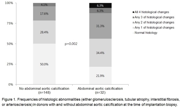
Results: Of 287 donors, 49 were stratified to AAC group and mean CS was 185.5 ± 263.3. AAC group was older than non-AAC group (51.1 ± 6.1 vs. 37.9 ± 11 years, respectively, p<0.001). Among AAC group, donors with CS>100 were associated with delayed recovery of renal function (p=0.035). In multivariable logistic regression, CS>100 (OR=12.4, p=0.017) and preoperative GFR (OR=0.829, p=0.001) were associated with delayed recovery of renal function. AAC group were more likely to have any histological abnormalities on implantation biopsies than non-AAC group (75.0% vs. 47.3%, p=0.004). Donors with AAC were more likely to have glomerulosclerosis (50.0% vs. 29.1%, p=0.022) and tubular atrophy (62.5% vs. 33.1%, p=0.002). Donors with AAC had a higher Nephrosclerosis score (p=0.002).
Conclusion: Living donors with CS >100 require close observation because they have a greater possibility of developing delayed recovery of renal function after donation. The presence of AAC is associated with various degrees of nephrosclerosis in healthy adults.
To cite this abstract in AMA style:
Yoon Y, Choi K, Joo D, Huh K, Kim M, Kim S, Kim Y, Yang S, Han W. Impact of Abdominal Aortic Calcification in Living Kidney Donors [abstract]. Am J Transplant. 2015; 15 (suppl 3). https://atcmeetingabstracts.com/abstract/impact-of-abdominal-aortic-calcification-in-living-kidney-donors/. Accessed February 12, 2026.« Back to 2015 American Transplant Congress
