Endocapillary NK Cells Are Increased in Indication Renal Transplant Biopsies with Antibody and T Cell Mediated Rejection.
Pathology, University of Michigan Medical School, Ann Arbor, MI
Meeting: 2017 American Transplant Congress
Abstract number: 301
Keywords: Alloantibodies, Kidney transplantation, Natural killer cells
Session Information
Session Name: Concurrent Session: Antibody Mediated Rejection in Kidney Transplant Recipients: Pathophsiology and Epidimiology
Session Type: Concurrent Session
Date: Monday, May 1, 2017
Session Time: 4:30pm-6:00pm
 Presentation Time: 5:30pm-5:42pm
Location: E354a
Detection of NK cell transcripts in renal transplant biopsies is associated with antibody mediated rejection (AMR). Similarly, quantification of endocapillary NK cells in paraffin tissue may be an alternative to the insensitive C4d assay in AMR diagnosis in conventional samples.
34 consecutive renal transplant indication biopsies were triple stained for CD16, Tbet, and CD34. Slides were digitized (Leica AT2) and cortical regions were analyzed using Imagescope. CD34 defined the peritubular capillaries and glomerular capillary loops. 2 blinded readers quantified CD16+/Tbet+ cells within capillary spaces.
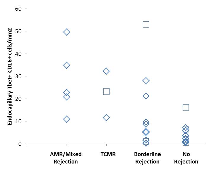
Most CD16+/Tbet+ cells were endovascular. Peritubular capillary CD16+/Tbet+ cells per mm2 cortex were elevated in biopsies with diagnoses of AMR (27.85+/-14.8) and pure T cell mediated rejection (TCMR, 22.4+/-10.4) compared to biopsies with non-rejection diagnoses (3.7+/-4.2, p<0.05 by t-test, figure 1). In addition, glomerular capillary CD16+/Tbet+ cells per mm2 glomerular area were also elevated in AMR (127+/-47) and TCMR (133+/-40) compared to no rejection (38+/-37, p<0.01). Biopsies with Borderline rejection had variable peritubular (12.4+/-16.1, p=0.06) and glomerular (52+/-47, p=0.41) capillary CD16+/Tbet+ cells per mm2. 3 patients with high and 7 patients with low CD16+/Tbet+ levels had or subsequently tested positive for donor specific antibodies (squares).
Endovascular CD16+/Tbet+ cells in biopsies meeting Banff criteria for AMR or mixed rejection is consistent with molecular data showing an NK cell signature of AMR. The elevated levels of CD16+/Tbet+ cells in cases of “pure” TCMR is unexpected, and we speculate that such cases may harbor non-HLA DSA or low levels of noncirculating DSA within the graft. In some biopsies called Borderline rejection but harboring high levels of CD16+/Tbet+ cells, review of histology confirms a subtle capillaritis that was not described in the pathology report. Immunohistochemical staining for NK cells has the potential to enhance the sensitivity of AMR diagnosis in standard paraffin embedded biopsies. Highlighting capillaries with CD34 aided manual counting, and may be useful in computer aided morphometry.
CITATION INFORMATION: Shammout A, Barnes J, Farkash E. Endocapillary NK Cells Are Increased in Indication Renal Transplant Biopsies with Antibody and T Cell Mediated Rejection. Am J Transplant. 2017;17 (suppl 3).
To cite this abstract in AMA style:
Shammout A, Barnes J, Farkash E. Endocapillary NK Cells Are Increased in Indication Renal Transplant Biopsies with Antibody and T Cell Mediated Rejection. [abstract]. Am J Transplant. 2017; 17 (suppl 3). https://atcmeetingabstracts.com/abstract/endocapillary-nk-cells-are-increased-in-indication-renal-transplant-biopsies-with-antibody-and-t-cell-mediated-rejection/. Accessed February 19, 2026.« Back to 2017 American Transplant Congress
