Association of Chest CT Score During Acute COVID-19 Illness with Subsequent Lung Function Loss Among Lung Transplant Patients
University of Texas Southwestern, Dallas, TX
Meeting: 2022 American Transplant Congress
Abstract number: 1485
Keywords: COVID-19, Morbidity, Radiologic assessment
Topic: Clinical Science » Lung » 64 - Lung: All Topics
Session Information
Session Time: 7:00pm-8:00pm
 Presentation Time: 7:00pm-8:00pm
Location: Hynes Halls C & D
*Purpose: Limited data exists on CT chest abnormalities during acute Coronavirus disease 2019 (COVID-19) infection and associated post-illness loss of lung function among lung transplant (LT) patients.
*Methods: The institutional database was interrogated for any LT patient diagnosed with COVID-19 during a one-year period (March 2020 to Feb 2021; n=54). 44 patients with acute COVID-19 were alive at 3-month follow up (COVID-survivors: 81.5%). Of the survivors, 34 had a CT chest during the first two weeks of acute illness. A validated CT score was used to quantify the parenchymal abnormalities due to COVID-19. Each lung was divided into 10 separate regions which were scored 0-2 based on the severity and extent of parenchymal opacification (maximum score per lung=20). To avoid confounding from underlying lung disease, only the allograph was assessed in single LT. The average score of both lungs was calculated in bilateral LT. The primary outcome measure was sustained decline of FEV1 or FVC >10% from pre-infection spirometry.
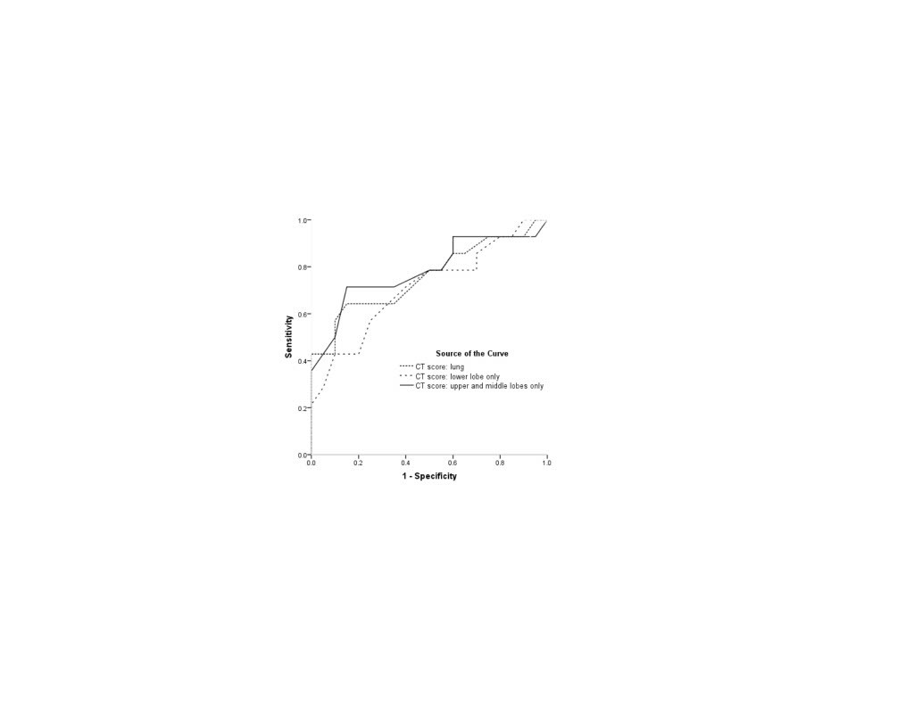
*Results: Abnormal CT score and lung opacities on CT chest were nearly ubiquitous during acute COVID-19 illness (>0; 36/37, 97.3%, median score with IDR: 7.25, 4.625-10.125). The lower lobes (LL) were more affected by COVID-19 than the upper and middle lobes (UML) (median CT score in LL: 4, 2.75-6 vs 3.5, 1.25-5 in UML). A >10% decline in FEV1 or FVC was common after COVID-19 pneumonia (38.2%). The overall CT score correlated with amount of lung function loss (r=0.36, p=0.03) although the association was modest and limited to regions reflecting the UML. On ROC curve, CT score was modestly predictive of lung function decline (Fig 1). CT score from UML had the highest area under the curve (78.2%, 61.1-95.4%; p=0.006) with a score of 4.5 being the best cut-off (sensitivity 71%, specificity 85% for post-COVID lung function loss >10%). An UML CT score >4.5 was strongly associated with respiratory failure during acute illness (69% vs 24%; OR: 7.2, 1.5-33.8; p=0.01) and lung function decline >10% (77% vs 19%; OR: 14.2, 2.6-76.7; p=0.001).
*Conclusions: The CT score during acute COVID-19 infection provides prognostic information regarding loss of lung function among LT patients who survive COVID-19. Parenchymal abnormalities in the UML best predict subsequent lung function loss.
To cite this abstract in AMA style:
Halverson QM, Batra K, Mahan LD, Mohanka M, Lawrence AC, Joerns J, Bollineni S, Kaza V, Timofte IL, Kershaw CD, Terada LC, Torres F, Banga A. Association of Chest CT Score During Acute COVID-19 Illness with Subsequent Lung Function Loss Among Lung Transplant Patients [abstract]. Am J Transplant. 2022; 22 (suppl 3). https://atcmeetingabstracts.com/abstract/association-of-chest-ct-score-during-acute-covid-19-illness-with-subsequent-lung-function-loss-among-lung-transplant-patients/. Accessed February 23, 2026.« Back to 2022 American Transplant Congress

