An Interesting Case of Proteinuria in Living Kidney Donor
A. Yadav, W. Maley, P. Singh
Thomas Jefferson University, Philadelphia, PA
Meeting: 2019 American Transplant Congress
Abstract number: B284
Keywords: Donors, unrelated, Kidney, Proteinuria, Screening
Session Information
Session Name: Poster Session B: Kidney Living Donor: Selection
Session Type: Poster Session
Date: Sunday, June 2, 2019
Session Time: 6:00pm-7:00pm
 Presentation Time: 6:00pm-7:00pm
Location: Hall C & D
*Purpose: Evaluation for proteinuria is vital to assess suitability for live kidney donation. Living unrelated donor with orthostatic proteinuria was evaluated. This case highlights importance of considering rare etiologies for orthostatic proteinuria, before potential donors are ruled out.
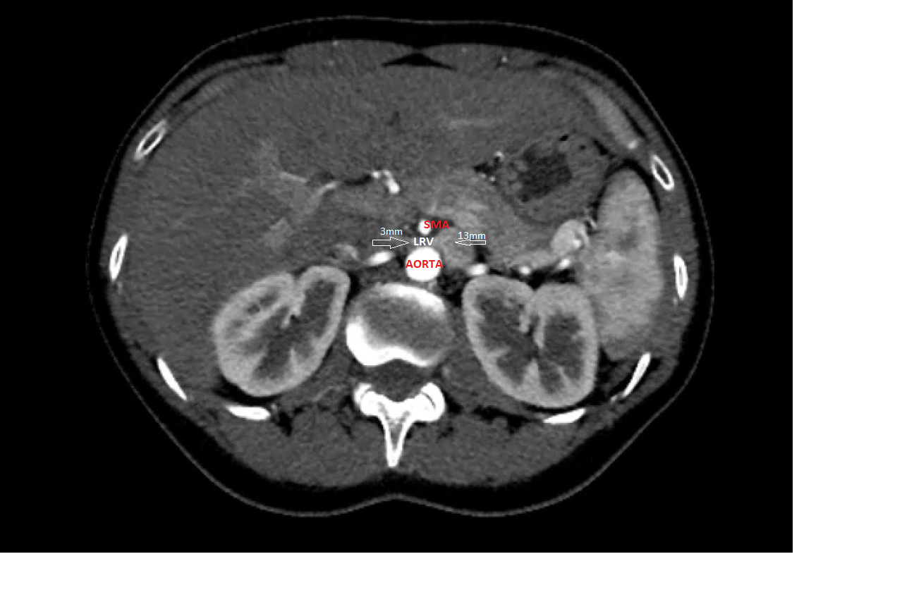
*Methods: A 41-year-old Caucasian female, was evaluated to be a live kidney donor to her husband who was on renal replacement therapy. She had no significant past medical or surgical history. Specifically, there was no history of urinary tract infection, nephrolithiasis, NSAIDs, herbal or protein supplements usage. Family history was non-contributory. On exam, she was normotensive with a BMI of 24. She possessed excellent renal function but demonstrated proteinuria >300 mg/24 hours on multiple occasions (Table 1). She had no hematuria and had negative urine analysis and culture. Split urine collection revealed orthostatic nature of this proteinuria with supine proteinuria of 50mg/8hr and upright proteinuria of 240mg/16hr (Table 1). Renal ultrasound showed normal sized kidneys with no hydronephrosis, stones or masses. Further serologic testing for Hepatitis B and C, Quantiferon and HIV were negative. ANA panel showed speckled pattern 1:40 titer with normal complement levels and normal serum and urine protein electrophoresis. Computed tomography revealed marked change in the caliber of left renal vein (LRV) from 13 mm to 3 mm as it passed behind the superior mesenteric artery. There was collateral vein extending posteriorly from the proximal vein (Image 1).
*Results: The anatomic findings represent nutcracker phenomenon (NCP) which predisposed this potential donor to orthostatic proteinuria. NCP is characterized by compression of LRV between superior mesenteric artery (SMA) and abdominal aorta. After informed consent specific to this scenario, she underwent left donor nephrectomy via laparoscopic approach with no complications. Six months post donation of kidney affected by NCP, complete resolution of proteinuria was noted in the donor. Additionally, the recipient has excellent allograft function with Serum Cr 1.0 mg/dl with no proteinuria.
*Conclusions: In summary, anatomic causes of proteinuria should be considered and imaging testing should be considered to ascertain such rare etiologies before potential donors are ruled out .
| Timeline | 24hr Creatinine clearance (ml/min) | Protein/24hr |
| 2 months prior to donation | 108 | 318 mg/24hr |
| 1 month prior to donation | 108 | 318 mg/24hr |
| 3 weeks prior to donation | Orthostatic split proteinuria: Supine:50mg/8hr Standing: 240mg/16hr | |
| Post donation | <34mg/24hr; Serum creatinine 1.0mg/dl |
To cite this abstract in AMA style:
Yadav A, Maley W, Singh P. An Interesting Case of Proteinuria in Living Kidney Donor [abstract]. Am J Transplant. 2019; 19 (suppl 3). https://atcmeetingabstracts.com/abstract/an-interesting-case-of-proteinuria-in-living-kidney-donor/. Accessed February 15, 2026.« Back to 2019 American Transplant Congress

