Alterations in Left Ventricular Wall Thickness after Cardiac Transplantation
Advanced Heart Failure, Transplant and Mechanical Circulatory Support, Montefiore Medical Center, Albert Einstein, Bronx, NY
Meeting: 2020 American Transplant Congress
Abstract number: B-266
Keywords: Multivariate analysis, N/A, Rejection, Ultrasonography
Session Information
Session Name: Poster Session B: Heart and VADs: All Topics
Session Type: Poster Session
Date: Saturday, May 30, 2020
Session Time: 3:15pm-4:00pm
 Presentation Time: 3:30pm-4:00pm
Location: Virtual
*Purpose: We investigated changes in cardiac wall thickness of donor hearts over time, including the peri-operative OHT period (within 4 days after transplantation) and up to 6 months post-operatively. In addition, we also sought to assess which clinical factors are associated with these changes over time, including early acute cellular rejection (ACR).
*Methods: This was a single center retrospective review of 100 consecutive OHT patients between May 2015 and May 2018 at our institution. Transthoracic echocardiographic (TTE) left ventricular (LV) wall thickness parameter measurements were obtained for donor hearts pre, peri (within 4 days after OHT) and as well as 6 months post transplantation. We also sought to analyze the association of clinical parameters and acute cellular rejection (ACR) on wall thickness over time by using multivariable linear regression.
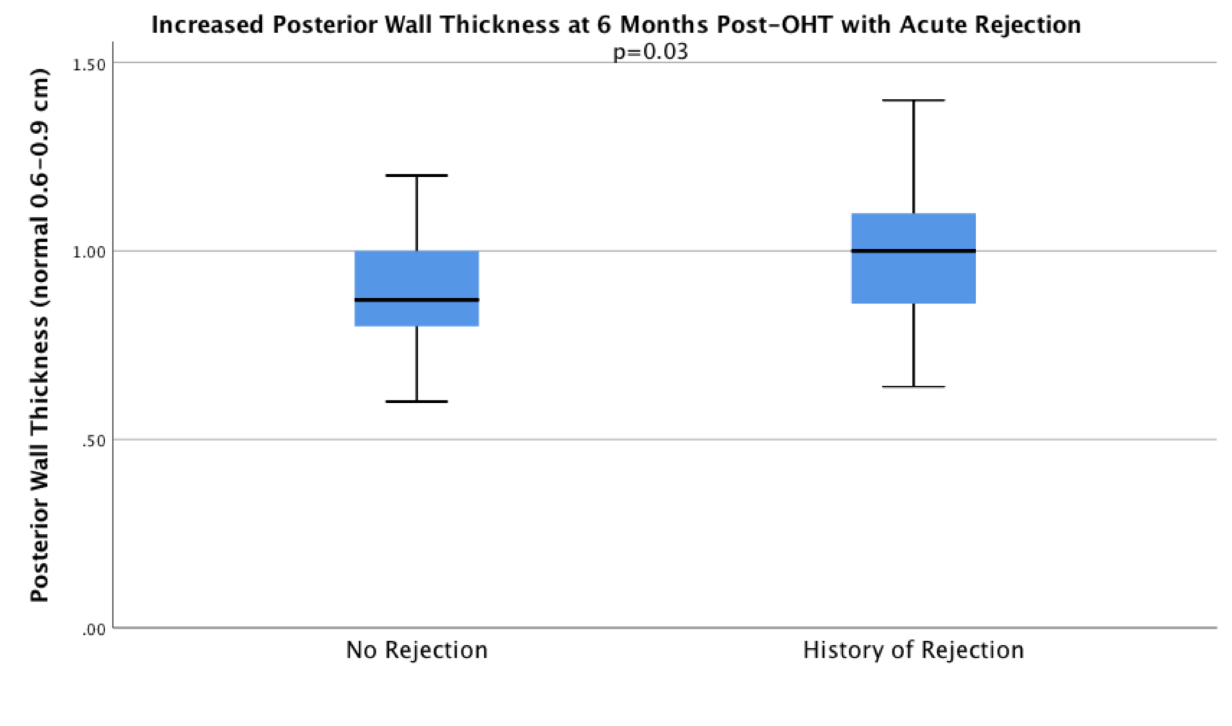
*Results: In the overall cohort, there was 60% male donors that were 34±12 years old, 72% male recipients that were 55±10 years old. Ischemic time was 202±49 minutes, and 15% had a female to male gender mismatch. LV posterior wall thickness (PWT) increased from 0.95cm±0.20 to 0.99cm±0.17, p<0.05, interventricular septum wall thickness (IVSWT) increased from 0.94cm±0.20 to 1cm±0.18, p<0.006, and relative wall thickness (RWT) also increased (0.44cm±0.11 to 0.49cm±0.24, p<0.03) from pre to peri-transplantation. At six months post OHT, wall thickness decreased (PWT: 0.92cm±0.15, p<0.0001, IVSWT: 0.94cm±0.16, p<0.001, RWT 0.42cm±0.10, p<0.005). There was no significant change in the mean left ventricular internal dimension at end diastole and at end systole from pre-OHT to 6 months post-OHT. Early ACR (beta: 0.077, p=0.03, figure 1), recipient history of hypertension (beta: 0.071, p= 0.018), and older recipient (beta: 0.003, p=0.005) was independently associated with a higher PWT and RWT at 6 months. In addition, increased peri-OHT PWT increased the risk of ACR (p=0.02).
*Conclusions: The initial increase in peri-operative LV wall thickness regresses by 6 months post-OHT. However, early acute cellular rejection, recipient history of hypertension, and increased recipient age are associated with a greater wall thickness at 6 months post-OHT.
To cite this abstract in AMA style:
Alvarez CK, Saeed O, Patel SR, Borukhov E, Jorde U. Alterations in Left Ventricular Wall Thickness after Cardiac Transplantation [abstract]. Am J Transplant. 2020; 20 (suppl 3). https://atcmeetingabstracts.com/abstract/alterations-in-left-ventricular-wall-thickness-after-cardiac-transplantation/. Accessed February 8, 2026.« Back to 2020 American Transplant Congress

